Oropouche virus, an emerging arbovirus that has swept through Central and South America in recent years, has long been viewed as a cause of unpleasant but self-limiting febrile illness. That perception is changing. Since 2023, large-scale outbreaks involving tens of thousands of confirmed cases have been reported, including in regions with no prior evidence of viral circulation, and clinicians have documented an increasing number of severe neurological complications, fatal outcomes, and adverse pregnancy events. Despite this escalating public health threat, no approved antiviral therapies or vaccines exist, and patient care remains entirely supportive. A new study published in PLOS Pathogens by Cássia Sousa Moraes, Yukari Itakura, and colleagues at Hokkaido University now offers the most detailed preclinical picture yet of how Oropouche virus disease unfolds—and demonstrates that an existing, licensed antiviral drug can halt it.
The drug in question is favipiravir, a broad-spectrum nucleoside analogue that inhibits viral RNA-dependent RNA polymerases and is already approved for influenza treatment in several countries. The research team began by characterizing its activity against Oropouche virus in cultured Vero E6 cells, comparing it directly with ribavirin, another broad-spectrum antiviral. Favipiravir proved substantially more potent, with a mean half-maximal effective concentration of 28.4 micromolar compared with 115.7 micromolar for ribavirin. Untreated infected cells developed marked cytopathic effects, and ribavirin offered only limited protection, whereas favipiravir preserved cellular morphology in a dose-dependent fashion and robustly reduced infectious virus production. Time-of-addition experiments pinpointed the drug’s stage of action: treatment significantly reduced viral titers when administered at the time of infection or during early replication, but not at the maturation stage, consistent with favipiravir’s established mechanism of interfering with viral RNA synthesis during the post-entry replication phase.
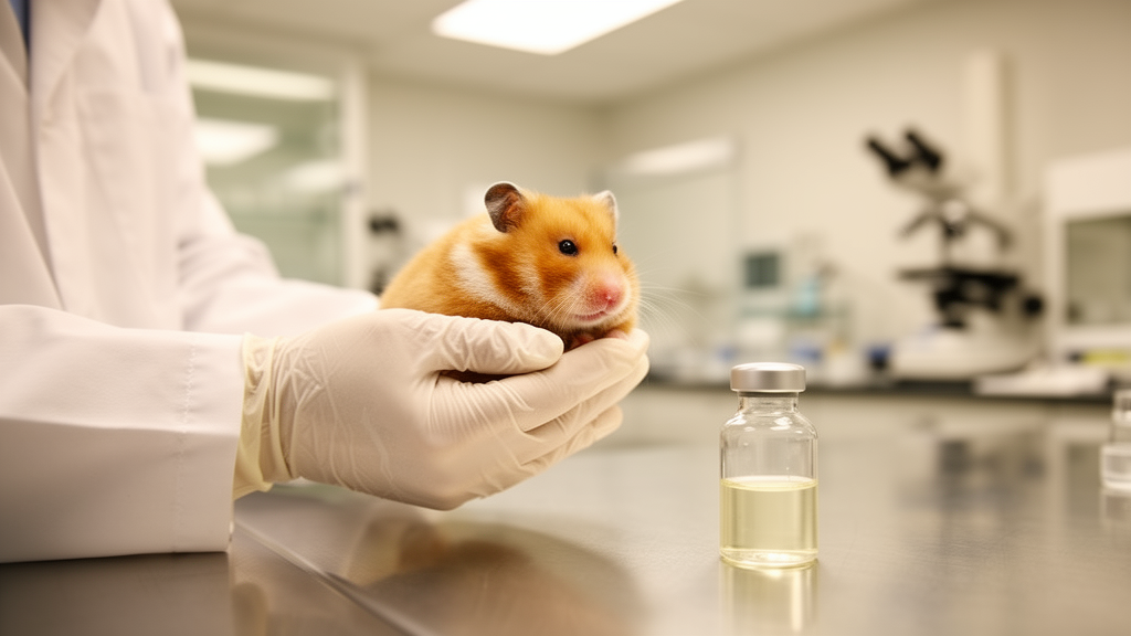
The decisive test came in vivo. The researchers used a lethal Syrian hamster model, infecting animals intraperitoneally with 5.4 × 10⁵ plaque-forming units of the OROV TRVL 9760 strain. All vehicle-treated animals succumbed to infection by three days post-infection, without preceding overt clinical symptoms or weight loss—a fulminant course that underscores how aggressive lethal Oropouche disease can be in this model. In stark contrast, animals receiving high-dose favipiravir at 600 milligrams per kilogram per day, given orally twice daily beginning one hour before infection and continuing for six days, survived the entire 26-day observation period without any clinical signs and steadily gained weight. Lower doses of favipiravir and both tested doses of ribavirin conferred only partial protection, and notably, some animals in the ribavirin groups developed neurological symptoms, including ataxia, hindlimb weakness, and paralysis, after treatment ceased.
Virological measurements explained these divergent outcomes. Using plaque assays and reverse transcription quantitative PCR, the team tracked viral loads in serum, brain, liver, and spleen at multiple time points. Vehicle-treated animals carried high viral burdens in all tissues at two days post-infection. High-dose favipiravir, by contrast, suppressed viral replication so completely that no viral RNA or infectious virus was detectable in any tissue at any time point examined. The suboptimal regimens told a more cautionary story: although low-dose favipiravir and ribavirin reduced infectious virus in serum, liver, and spleen, infectious virus persisted in the brains of these animals at six and eight days post-infection, indicating incomplete clearance from the central nervous system that correlated with the delayed neurological disease observed after treatment withdrawal. The message is clear—insufficient antiviral pressure allows viral neuroinvasion, and neuroinvasion drives fatal disease.
Perhaps the most clinically encouraging finding concerns timing. In outbreak settings, patients rarely present before infection is already established, so a drug that works only as prophylaxis would be of limited value. The researchers therefore tested delayed treatment, initiating favipiravir at one hour before infection, at the time of infection, or at 6, 24, or 48 hours after infection. Strikingly, animals that began treatment as late as 24 hours post-infection were fully protected, with 100 percent survival and complete suppression of viral replication in systemic organs and the central nervous system. Even at 48 hours post-infection, when the therapeutic window was narrowing, favipiravir still conferred partial protection, with 50 percent survival and consistent weight gain among survivors. This therapeutic window, demonstrated in a model where untreated infection is uniformly lethal within days, substantially strengthens the case for favipiravir as a practical candidate for human Oropouche disease.
Beyond efficacy, the study’s second major contribution is mechanistic. To define the host responses associated with Oropouche virus pathogenesis and their modulation by antiviral therapy, the team performed tissue-resolved transcriptomic profiling—RNA sequencing of liver and brain collected at two days post-infection. Because the Syrian hamster genome remains less comprehensively annotated than the mouse genome, hamster genes were functionally annotated through their mouse orthologs. Principal component analysis revealed a clear transcriptional separation between infected and uninfected animals in both tissues, while favipiravir-treated animals clustered near uninfected controls, indicating that the infection-driven transcriptional upheaval was largely suppressed by the drug. This is, to the authors’ knowledge, the first in vivo, tissue-resolved transcriptomic framework of Oropouche virus infection.
The transcriptomic signatures in infected animals were distinctive and, in places, ominous. In the liver, where the virus replicated robustly, infection induced upregulation of genes involved in chemotaxis, cytokine signaling, myeloid cell differentiation, and interferon responses, alongside downregulation of metabolic pathways governing energy production and fatty acid metabolism. In the brain, antiviral and inflammatory programs were induced even more strongly, accompanied by pathways promoting immune cell recruitment. Quantitative PCR validation of selected interferon-stimulated genes—Cxcl10, Rsad2, and Isg15—confirmed strong induction at two days post-infection in both tissues of untreated animals, which was suppressed by both favipiravir and ribavirin at that early stage. At later time points, however, Rsad2 and Isg15 expression tended to remain elevated in ribavirin-treated animals, correlating with their persistent brain viral loads, whereas expression stayed low in favipiravir-treated animals. Interestingly, the canonical inflammatory cytokines Il6, Il10, and Tnf-alpha showed no significant changes at two days post-infection and did not track with viral replication, suggesting they are unlikely to be major drivers of Oropouche disease in this model.
The metabolic findings deserve particular attention. Although liver involvement is not a prominent feature of typical Oropouche fever, recent fatal human infections have been associated with markedly elevated aminotransferase levels and liver injury biomarkers, and independent studies have demonstrated hepatic infection and inflammatory responses in human liver organoids and an experimental mouse model. The simultaneous enrichment of pathways associated with positive regulation of innate immune responses and negative regulation of defense responses in high-burden animals suggests a complex, potentially dysregulated host response, in which sustained antiviral and inflammatory signaling coexists with regulatory mechanisms that constrain excessive immune activation. When viral replication remains high, such regulation appears insufficient. The concurrent downregulation of energetic, lipid metabolic, and mitochondrial pathways in the liver implies that cellular stress during high-burden infection may contribute to tissue dysfunction and injury, meaning that Oropouche pathogenesis likely reflects not only direct viral replication but also the interplay between persistent viral burden, dysregulated immunity, and disrupted metabolic homeostasis.
The authors are candid about the study’s limitations. The reliance on mouse orthologs for gene annotation means hamster-specific transcriptional features and genes lacking confident mouse orthologs may have been underrepresented. Additionally, because all vehicle-treated animals reached the humane endpoint before six days post-infection, direct comparisons of gene expression between untreated and treated animals at later time points were impossible, leaving some uncertainty about whether the later profiles reflect favipiravir’s effects or the absence of ongoing infection. These caveats notwithstanding, the study extends earlier work by Rodrigues and colleagues, who established the Syrian hamster as a relevant model of Oropouche pathogenesis, by integrating tissue-resolved transcriptomics with therapeutic intervention in a single experimental framework.
Translational considerations will shape what happens next. Favipiravir offers genuine advantages over other polymerase-targeting candidates such as 4′-fluorouridine, including oral bioavailability and prior clinical use against multiple viral diseases, and its demonstrated efficacy with delayed initiation enhances its relevance for outbreak settings where early diagnosis is difficult. Yet the drug carries known safety liabilities: teratogenic and embryotoxic effects, and an association with elevated serum uric acid and hyperuricemia. These concerns are especially pertinent for Oropouche virus, given the growing recognition of congenital and pregnancy-associated disease. The authors emphasize that further studies addressing pharmacokinetics, optimal dosing, treatment duration, and safety in relevant populations will be required before clinical application. Even so, the findings provide strong preclinical evidence that timely, potent antiviral intervention can prevent central nervous system invasion, suppress pathogenic inflammatory and metabolic programs, and avert lethal Oropouche disease—transforming the outlook for a pathogen that, until now, medicine could only watch.
Subject of Research: Favipiravir efficacy and host transcriptomic responses in lethal Oropouche virus infection
Article Title: Integrated in vivo and transcriptomic analyses of lethal Oropouche virus infection reveal suppression of pathogenic host responses by antiviral therapy
Article References: Sousa Moraes, C., Gonzalez, G., Sato, A., Miki, S., Inoue, A., Tabata, K., Kranrod, J. W., Kabamba, C. F., Ohnuma, A., Matsuno, K., Harada, R., Saito, S., Akimoto, N., Sasaki, M., Orba, Y., Hall, W. W., Sawa, H., & Itakura, Y. (2026). Integrated in vivo and transcriptomic analyses of lethal Oropouche virus infection reveal suppression of pathogenic host responses by antiviral therapy. PLOS Pathogens, 22(10), e1014647. https://doi.org/10.1371/journal.ppat.1014647
Image Credits: AI Generated
DOI: 10.1371/journal.ppat.1014647
Keywords: Oropouche virus, favipiravir, antiviral therapy, arbovirus, neuroinvasion, Syrian hamster model, transcriptomics, interferon response, ribavirin, PLOS Pathogens, emerging infectious disease, RNA virus
News Source: Kristina Jarvis. (October 8, 2026). Antiviral Drug Favipiravir Blocks Lethal Oropouche Virus Infection in Hamsters. Scienmag.