• HOME
  • NEWS
  • EXPLORE
    • CAREER
      • Companies
      • Jobs
    • EVENTS
    • iGEM
      • News
      • Team
    • PHOTOS
    • VIDEO
    • WIKI
  • BLOG
  • COMMUNITY
    • FACEBOOK
    • INSTAGRAM
    • TWITTER
Thursday, May 21, 2026
BIOENGINEER.ORG
No Result
View All Result
  • Login
  • HOME
  • NEWS
  • EXPLORE
    • CAREER
      • Companies
      • Jobs
        • Lecturer
        • PhD Studentship
        • Postdoc
        • Research Assistant
    • EVENTS
    • iGEM
      • News
      • Team
    • PHOTOS
    • VIDEO
    • WIKI
  • BLOG
  • COMMUNITY
    • FACEBOOK
    • INSTAGRAM
    • TWITTER
No Result
View All Result
Bioengineer.org
No Result
View All Result
Home NEWS Science News Health

High-speed whole-brain imaging improves understanding of brain disease

Bioengineer by Bioengineer
July 10, 2017
in Health
Reading Time: 2 mins read
0
Share on FacebookShare on TwitterShare on LinkedinShare on RedditShare on Telegram
IMAGE

Credit: Osaka University

Osaka, Japan – To fully understand brain function and dysfunction, it is important to be able to visualize changes in anatomy and activity in the whole brain. High-resolution brain imaging that can distinguish individual cells and quantitative comparison of acquired data are essential to show how the brain is affected by disease.

However, current attempts to image a whole mouse brain at a resolution high enough to gain detailed information take up to one week. While these approaches have revealed important insights into brain function, it is not possible to image and analyze multiple brains with these technologies. Comparing multiple brains is essential to understand neurobiological function and dysfunction in brain disorders.

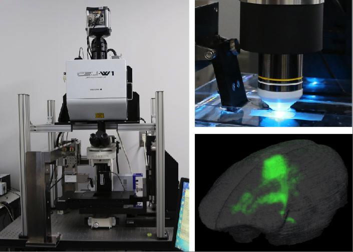
Osaka University-led researchers have developed block-face serial microscopy tomography (FAST)–an imaging system that can image a whole mouse brain at high spatial resolution in less than two-and-a-half hours. "FAST consists of a spinning disk confocal microscope with built in microslicer and a method for processing image data," explains first author Kaoru Seiriki. "With our 3D reconstruction technique, whole brains can be visualized at a resolution high enough to resolve individual cells and their subcellular structures."

By combining their FAST technique with specific staining procedures, Seiriki and colleagues were able to visualize subcellular nuclei, vascular structures, mature oligodendrocytes, myelin sheaths, interneurons, and projecting neurons throughout the whole brain. These imaging tools provide a systemic approach to investigating the pathophysiological mechanisms of different brain diseases.

FAST is a very quick imaging technique, therefore it can potentially be used to image non-human primate brains. "We successfully visualized a long-range neuronal projection at a subcellular resolution in the whole brain of an adult marmoset," says leading researcher Hitoshi Hashimoto. "This shows how FAST can further our understanding of brain anatomy in rodents and primates."

The Osaka University research team has also successfully imaged postmortem human brains using their FAST system. "We expect that this approach will identify fine morphological abnormalities in diseased human brains that were previously unknown," says Hashimoto. These insights may help to advance the development of effective treatments.

This new technique offers a way to compare multiple brains at the level of individual cells and their subcellular structures. With this approach, new insights will be gained into the pathological mechanisms of different brain diseases. Furthermore, the applicability of this offers a translational approach to researching non-human animal models and human diseases.

###

Media Contact

Saori Obayashi
[email protected]
81-661-055-886
@osaka_univ_e

http://www.osaka-u.ac.jp/en

Original Source

http://www.cell.com/neuron/abstract/S0896-6273(17)30455-5 http://dx.doi.org/10.1016/j.neuron.2017.05.017

Share12Tweet8Share2ShareShareShare2

Related Posts

Genetic Insights from 619,372 Metabolic Profiles

May 21, 2026

Bacterial STIs Hit Record Levels in Europe as Congenital Syphilis Cases Nearly Double

May 21, 2026

Oral Semaglutide Lowers Cardiometabolic Risks in Obesity

May 21, 2026

Nomogram Predicts 30-Day Mortality in Elderly HLH

May 21, 2026
Please login to join discussion

POPULAR NEWS

  • blank

    New Study Reveals Plants Can Detect the Sound of Rain

    733 shares
    Share 292 Tweet 183
  • ESMO 2025: mRNA COVID Vaccines Enhance Efficacy of Cancer Immunotherapy

    304 shares
    Share 122 Tweet 76
  • Research Indicates Potential Connection Between Prenatal Medication Exposure and Elevated Autism Risk

    846 shares
    Share 338 Tweet 212
  • Breastmilk Balances E. coli and Beneficial Bacteria in Infant Gut Microbiomes

    58 shares
    Share 23 Tweet 15

About

We bring you the latest biotechnology news from best research centers and universities around the world. Check our website.

Follow us

Recent News

Label-Free Super-Resolution Imaging of Live Cells

Genetic Insights from 619,372 Metabolic Profiles

Bacterial STIs Hit Record Levels in Europe as Congenital Syphilis Cases Nearly Double

Subscribe to Blog via Email

Enter your email address to subscribe to this blog and receive notifications of new posts by email.

Join 82 other subscribers
  • Contact Us

Bioengineer.org © Copyright 2023 All Rights Reserved.

Welcome Back!

Login to your account below

Forgotten Password?

Retrieve your password

Please enter your username or email address to reset your password.

Log In
No Result
View All Result
  • Homepages
    • Home Page 1
    • Home Page 2
  • News
  • National
  • Business
  • Health
  • Lifestyle
  • Science

Bioengineer.org © Copyright 2023 All Rights Reserved.