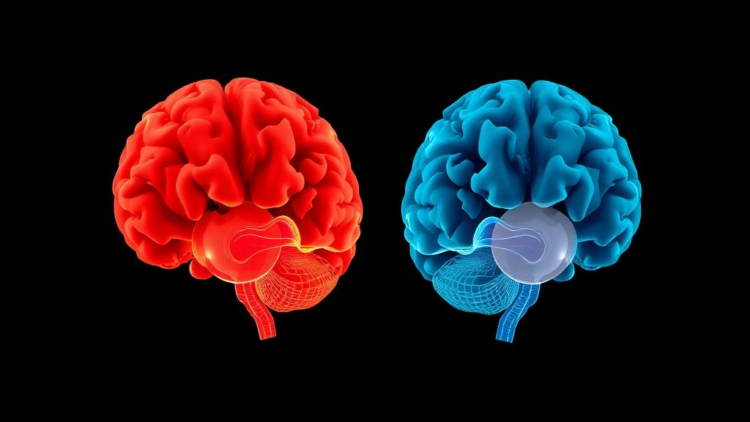
For decades, neuroscientists have marveled at one of the brain’s most striking tricks: when hearing is lost early in life, the auditory cortex does not fall silent. Instead, it is commandeered by the surviving senses, particularly vision, and put to work on entirely new jobs. But a fundamental question has divided the field. Does the deprived hearing cortex genuinely change its function, adopting wholly new cognitive roles, or does it preserve its original computational specialty, simply applying it to whatever input it can get? A new meta-analysis, the largest of its kind, now delivers a quantitative answer: both things happen at once, and the brain keeps them neatly separated across neighboring patches of cortex.
The study, published in the journal iScience, was led by Xiaoyi Zuo, Gantang Li, Zhengye Wang, Peng Peng, Chenjie Dong, and Suiping Wang, who pooled neuroimaging data from forty-seven studies comprising 801 deaf participants and 880 hearing controls. Most of the deaf participants had severe-to-profound hearing loss, 88 percent had lost hearing before the age of three, and 91 percent were sign language users. Rather than relying on qualitative narrative reviews, which have struggled to reconcile heterogeneous experimental paradigms, brain morphology, and activation patterns across studies, the team used activation likelihood estimation, or ALE, a coordinate-based meta-analytic technique that models each reported brain activation peak as a three-dimensional Gaussian probability distribution and tests whether activation converges across experiments more than chance would predict.
The first finding confirmed what single studies had long suggested: across all forty-seven studies, deaf participants consistently showed greater activation than hearing participants in the bilateral superior temporal gyri, the cortical ridge on each side of the brain that houses the primary and association auditory areas. Crucially, the researchers did not simply trust anatomical landmarks, a common shortcut in this literature that only about 11 percent of prior studies avoided. Instead, they defined the auditory cortex functionally, using auditory task activations in hearing participants combined with large-scale functional masks from the Neurosynth database. The result was striking: 82 percent of the voxels that lit up cross-modally in deaf participants fell within the functional auditory mask, confirming that the recruitment genuinely targets the deprived hearing cortex rather than adjacent, unrelated tissue.
The heart of the study lay in splitting the literature into two categories. Linguistic tasks, such as sign language comprehension and lipreading, made up 49 percent of the studies and probe domain-specific language processing. Non-linguistic tasks, such as tracking moving dots or judging faces, made up 38 percent and probe a broad range of visual processes without any linguistic content. If the functional change principle governed cross-modal plasticity, both task types should recruit the same auditory subregions. If functional preservation ruled, the two task types should engage distinct subregions. The data delivered a third, hybrid answer that supports a differential model of cross-modal plasticity.
Contrast analyses revealed that the left anterior superior temporal gyrus was recruited selectively during linguistic tasks. This region is a core node of the left-lateralized language network in hearing people, and its exclusive engagement by sign language and lipreading in deaf individuals suggests that its linguistic specialization survives auditory deprivation intact, merely switching its input channel from sound to sight. The laterality analysis reinforced this interpretation: linguistic tasks produced significantly left-lateralized cross-modal recruitment, with an average laterality index of 0.28, mirroring the left-hemisphere dominance for language seen in hearing brains. In other words, the auditory cortex of a deaf signer appears to keep doing language, just in a visual language.
In contrast, the left posterior superior temporal gyrus and the right superior temporal gyrus were recruited by both linguistic and non-linguistic tasks, and non-linguistic tasks alone produced strongly right-lateralized recruitment, with a laterality index of minus 0.75. These regions appear to have undergone a genuine functional shift toward domain-general visual processing, handling everything from object matching and motion perception to spatial attention and visual working memory. The researchers suggest this versatility may reflect the multifunctional profile of these regions in hearing people, where the right superior temporal gyrus contributes to acoustic-phonetic encoding, prosody perception, and spatial processing alike, giving it a flexibility that lends itself to repurposing when sound disappears.
The team went to unusual lengths to rule out confounds, running five separate control analyses. Because sign language and lipreading involve dynamic visual stimuli while many non-linguistic tasks use static images, they repeated the analysis restricted to dynamic stimuli and found the same pattern. They reclassified studies by task demands rather than stimulus type, again with the same result. Restricting the dataset to prelingual deafness, defined as hearing loss before age three, also replicated the findings. A leave-one-out jackknife analysis showed the clusters were robust to the exclusion of any single study, with an average robustness of 99 percent, and fail-safe N analyses indicated the results could withstand the addition of substantial numbers of unpublished null studies. The lone exception was reduced activation in the left inferior occipital gyrus, which showed susceptibility to publication bias and should be treated cautiously.
Parallels with blindness make the pattern more compelling. In blind individuals, the fusiform face area and the visual word form area preserve their specializations through touch or sound, while the primary visual cortex becomes a domain-general workhorse recruited for Braille reading, speech processing, and even mathematics. The deaf brain appears to follow the same logic in reverse: some temporal regions act as scaffolds that keep their original jobs through alternative senses, while others become flexible generalists. The authors note that preserved functions tend to be those that naturally operate across multiple senses, such as identity recognition and rhythm processing, which may be maintained by strengthening or unmasking pre-existing connections between auditory regions and multisensory cortices.
The findings carry real clinical weight. Cross-modal recruitment is known to influence outcomes after cochlear implantation, and the new framework offers a way to predict which patients might benefit. One hypothesis is that functionally preserved regions, such as the left anterior superior temporal gyrus, act as scaffolds that support auditory and speech rehabilitation, and activity in some cross-modally recruited areas has indeed been found to positively predict auditory outcomes. Conversely, regions that have undergone functional change toward domain-general visual processing may be associated with maladaptive outcomes, suggesting that targeted auditory training tailored to specific functions may be needed to restore their original roles. Longitudinal studies tracking cross-modal recruitment before and after implantation are the logical next step.
The authors also acknowledge limits. Coordinate-based meta-analyses of fMRI data identify converging BOLD signals but cannot map the fine-grained computational architecture underneath, and the broad linguistic versus non-linguistic taxonomy may partly reflect differences in attentional demand or cognitive load rather than pure domain specificity. The control analysis for dynamic non-linguistic tasks rested on only eleven experiments, below the recommended threshold of seventeen for robust ALE inference. Still, the central message stands and is likely to reshape how the field thinks about sensory deprivation: the deaf brain is neither a story of wholesale reorganization nor of rigid preservation, but a mosaic in which neighboring patches of the auditory cortex simultaneously hold onto language and reinvent themselves for vision, revealing an interplay between cortical architecture, sensory loss, and language experience that no single principle could capture alone.
Subject of Research: Cross-modal functional plasticity of the auditory cortex in deaf individuals
Article Title: Differential cross-modal functional plasticity in deaf individuals: A meta-analysis of neuroimaging studies
Article References: Zuo, X., Li, G., Wang, Z., Peng, P., Dong, C., & Wang, S. (2026). Differential cross-modal functional plasticity in deaf individuals: A meta-analysis of neuroimaging studies. iScience, 29(10), Article 117610. https://doi.org/10.1016/j.isci.2026.117610
Image Credits: AI Generated
DOI: 10.1016/j.isci.2026.117610
Keywords: cross-modal plasticity, deafness, auditory cortex, superior temporal gyrus, meta-analysis, neuroimaging, fMRI, sign language, brain plasticity, cochlear implantation, language processing, visual processing
Cite Scienmag News
APA
MLA
Chicago
Colin Clarke. (September 30, 2026). Deaf Brains Repurpose Sound Regions in Two Distinct Ways, Landmark Meta-Analysis Reveals. Scienmag. https://scienmag.com/deaf-brains-repurpose-sound-regions-in-two-distinct-ways-landmark-meta-analysis-reveals/
Colin Clarke. “Deaf Brains Repurpose Sound Regions in Two Distinct Ways, Landmark Meta-Analysis Reveals.” Scienmag, 30 September 2026, https://scienmag.com/deaf-brains-repurpose-sound-regions-in-two-distinct-ways-landmark-meta-analysis-reveals/. Accessed 30 September 2026.
Colin Clarke. “Deaf Brains Repurpose Sound Regions in Two Distinct Ways, Landmark Meta-Analysis Reveals.” Scienmag. September 30, 2026. https://scienmag.com/deaf-brains-repurpose-sound-regions-in-two-distinct-ways-landmark-meta-analysis-reveals/
Copy citation
Download RIS
Tags: auditory cortexauditory cortex reorganization in deaf individualsbrain plasticitycochlear implantationcross-modal neuroplasticity in sensory deprivationcross-modal plasticitydeafnessDeafness and brain plasticitydistinct neural adaptations in auditory cortexearly-onset deafness and brain functionfMRIfunctional repurposing of sound regions in the brainhemispheric specialization in sensory deprivationlanguage processingmeta-analysisneighboring cortical patches in sensory adaptationneuroimagingneuroimaging meta-analysis of deafnessneuroplasticity mechanisms in deaf populationssign languagesign language influence on brain reorganizationsuperior temporal gyrusvisual compensation in deaf brainsvisual processing