Triple-negative breast cancer has long been one of the most stubborn targets in oncology. Lacking the estrogen receptor, progesterone receptor, and HER2 amplification that anchor therapies for other breast cancer subtypes, it leaves patients with few options beyond chemotherapy and, more recently, immune checkpoint inhibitors. Yet even immunotherapy helps only a fraction of patients, and clinicians have struggled to predict who will benefit. A new study published in Clinical Proteomics offers a strikingly detailed answer to a deceptively simple question: when a triple-negative breast tumor looks inflamed under the microscope, where exactly is that inflammation located, and does it actually mean the immune system is fighting the cancer?
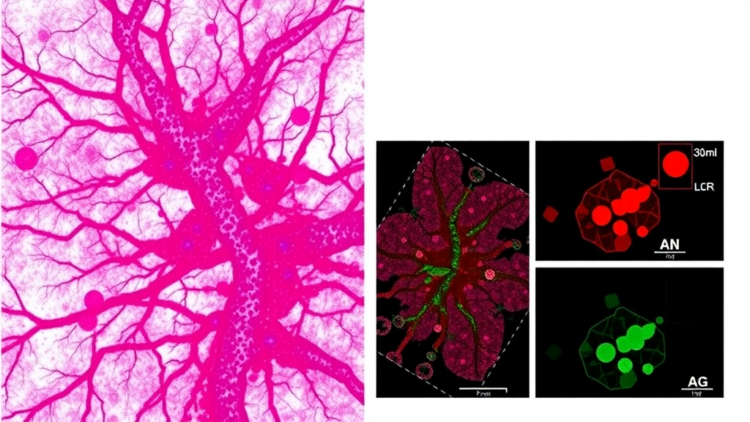
The research team, led by Allison L. Hunt and Thomas P. Conrads of the Inova Health System and Uniformed Services University together with Fei Wang and Yu Tian of AbbVie, took an unusually surgical approach to that question. Rather than grinding up whole tumor samples, the standard practice in most bulk molecular analyses, they used laser microdissection to physically separate two compartments within each tumor: the tumor epithelium, the malignant cells themselves, and the tumor-involved stromal microenvironment, the surrounding tissue of fibroblasts, immune cells, and blood vessels that shelters the cancer. Working with formalin-fixed paraffin-embedded, chemotherapy-naive specimens from twelve tumors obtained from eleven unique patients, they carved out these compartments with a laser beam guided by pathologist-annotated regions of interest.
The technical ambition of the study lies in what happened next. Each compartment was subjected to deep quantitative proteomic profiling using high-resolution liquid chromatography coupled to tandem mass spectrometry with data-independent acquisition, a strategy that systematically fragments and measures peptides across the entire sample rather than cherry-picking the most abundant ones. The result was a catalogue of 6,842 proteins co-quantified across every sample, an unusually deep measurement for microdissected archival tissue. This depth matters because proteins, not genes, are the functional machinery of cells, and measuring them directly captures the actual signaling states, metabolic programs, and inflammatory circuits operating inside each tumor compartment at the moment of surgical removal.
To classify tumors as immunologically hot or cold, the team began with pathology-assigned immune classifications, identifying six immune-hot and six immune-cold specimens based on hematoxylin and eosin stained sections, the routine stained slides pathologists examine every day. They then refined these assignments using ProteoMixture admixture deconvolution, a computational method that infers the composition of infiltrating immune cell populations from the protein signatures present in each sample. This dual approach, combining the trained eye of the pathologist with quantitative proteome deconvolution, allowed the researchers to anchor their molecular findings in histologic reality rather than treating each tumor as an undifferentiated blob of tissue.
The central finding is one of profound spatial compartmentalization. In immune-hot tumors, the stroma, not the tumor cells, carried the robust immunological signal. Stromal compartments displayed strong signatures of both innate immunity, the rapid, nonspecific first line of defense, and adaptive immunity, the antigen-specific arm involving T and B lymphocytes. The stromal profiles correlated with interferon signaling, antigen presentation machinery, and modules associated with tumor plasticity, all hallmarks of a tissue actively engaged in immune surveillance. In sharp contrast, the tumor epithelium from the same immune-hot specimens showed only weaker, predominantly innate inflammatory signaling, with heterogeneous metabolic and apoptotic signatures that varied considerably from patient to patient.
That asymmetry carries a sobering implication. The malignant cells themselves, even in tumors teeming with lymphocytes, appeared to be mounting at best a feeble inflammatory response, one that the authors suggest may be inadequate to overcome tumor-intrinsic immunosuppression or to robustly activate and engage adaptive immunity. In other words, the cancer cells were not cooperating with the immune cells camped at their borders. Immune infiltration, the study concludes, does not by itself predict effective antitumor immunity in triple-negative breast cancer. A tumor can look hot on a slide and still be functionally cold where it matters most, at the interface between the malignant cell and the immune system trying to destroy it.
The pathway-level analysis added further texture to this picture. The researchers integrated their proteomic data with gene module signatures derived from previously published single-cell RNA sequencing data from twenty-six breast cancers, including ten triple-negative tumors, allowing them to map protein-level changes onto cell-type-specific transcriptional programs. They found compartment-specific and immune-status-specific alterations in mTOR signaling, a central growth-control pathway. PI3K/AKT/mTORC1 signaling and its downstream metabolic programs were largely enriched in immune-hot stroma, suggesting that the inflamed stromal compartment is not merely a passive bystander but a metabolically active participant in the tumor ecosystem. Meanwhile, immune-cold tumors were enriched for mitotic and cell cycle progression pathways, consistent with a phenotype dominated by relentless proliferation rather than inflammatory crosstalk.
These findings arrive at a moment when the field is rethinking how it evaluates the immune response to solid tumors. Immune checkpoint inhibitors targeting PD-1 and PD-L1 have transformed treatment for a subset of triple-negative breast cancer patients, particularly those whose tumors express PD-L1 or carry high tumor-infiltrating lymphocyte counts. But the new data suggest that such histologic and single-marker assessments may be misleading. A stroma crowded with immune cells exhibiting interferon and antigen presentation signatures is not the same as a tumor epithelium that has surrendered to immune attack. Therapies, the authors argue, must target both the stromal immune dysfunction that prevents effective infiltration and activation, and the tumor-intrinsic resistance mechanisms that allow malignant cells to ignore or suppress whatever immune pressure does reach them.
The methodological lesson may prove as influential as the biological one. Laser microdissection of archival formalin-fixed paraffin-embedded tissue, combined with deep data-independent acquisition proteomics and computational immune deconvolution, transforms the humble pathology slide into a spatially resolved molecular map. Because FFPE blocks are stored in every hospital pathology department in the world, this workflow could be applied retroactively to thousands of tumors from completed clinical trials, asking retrospectively which patients responded to immunotherapy and how their tumor and stromal proteomes differed from those of nonresponders. The study was partially funded by AbbVie, and several authors are company employees, a detail that underscores the pharmaceutical industry’s growing interest in spatial proteomics as a tool for designing combination therapies that pair checkpoint inhibitors with agents targeting stromal signaling, metabolic reprogramming, or innate immune pathways such as NF-kappaB and toll-like receptor signaling.
For patients with triple-negative breast cancer, the immediate clinical impact of this study is not a new drug but a new map, one that shows immunologists precisely where the battle lines are drawn within a tumor. The stroma emerges as the true theater of the immune response, rich with innate and adaptive machinery, while the epithelial compartment harbors metabolic and cell-cycle programs that may explain why immune presence so often fails to become immune destruction. If future therapies can be engineered to convert that stromal inflammation into genuine epithelial engagement, perhaps by combining checkpoint blockade with agents that flip tumor cells from proliferative, immunologically silent programs into inflammatory, antigen-presenting ones, the promise of immunotherapy for this aggressive disease may finally extend beyond the fortunate few. The study is a reminder that in cancer biology, as in real estate, everything depends on location.
Subject of Research: Histology-resolved proteomic characterization of the tumor immune microenvironment in triple-negative breast cancer
Article Title: Histology resolved proteomic characterization of the triple negative breast cancer tumor immune microenvironment
Article References: Hunt, A. L., Wang, F., Abulez, T., Wang, X., Cui, B., Jin, L., Schejbal, J., Coberly, S., Ogata, J. D., Wilson, K. N., Conrads, K. A., Hood, B. L., Bateman, N. W., Uziel, T., Samayoa, J. A., Conrads, T. P., & Tian, Y. (2026). Histology resolved proteomic characterization of the triple negative breast cancer tumor immune microenvironment. Clinical Proteomics. https://doi.org/10.1186/s12014-026-09629-w
Image Credits: AI Generated
DOI: 10.1186/s12014-026-09629-w
Keywords: triple-negative breast cancer, tumor microenvironment, proteomics, laser microdissection, mass spectrometry, tumor stroma, immune checkpoint inhibitors, mTOR signaling, tumor-infiltrating lymphocytes, spatial proteomics, cancer immunotherapy, FFPE tissue
Cite Scienmag News
APA MLA Chicago
Kenneth Gardner. (September 26, 2026). Laser-Dissected Proteomics Reveals Where Immunity Really Lives in Triple-Negative Breast Tumors. Scienmag. https://scienmag.com/laser-dissected-proteomics-reveals-where-immunity-really-lives-in-triple-negative-breast-tumors/
Kenneth Gardner. “Laser-Dissected Proteomics Reveals Where Immunity Really Lives in Triple-Negative Breast Tumors.” Scienmag, 26 September 2026, https://scienmag.com/laser-dissected-proteomics-reveals-where-immunity-really-lives-in-triple-negative-breast-tumors/. Accessed 26 September 2026.
Kenneth Gardner. “Laser-Dissected Proteomics Reveals Where Immunity Really Lives in Triple-Negative Breast Tumors.” Scienmag. September 26, 2026. https://scienmag.com/laser-dissected-proteomics-reveals-where-immunity-really-lives-in-triple-negative-breast-tumors/
Copy citation Download RIS
Tags: cancer immune infiltrationcancer immunotherapyFFPE tissueimmune checkpoint inhibitorsimmune response in breast cancerimmunotherapy predictionlaser microdissectionmass spectrometrymTOR signalingProteomicsproteomics analysisspatial proteomicsstromal microenvironmenttargeted cancer therapiestriple-negative breast cancertumor epitheliumtumor heterogeneitytumor inflammation localizationtumor microenvironmenttumor stromatumor-infiltrating lymphocytes