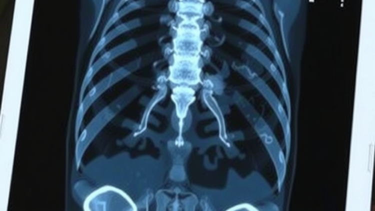
Medical images are often difficult to read, but few imaging modalities test the limits of human and machine perception quite like functional nuclear medicine. Single-photon emission computed tomography, or SPECT, bone scintigraphy produces whole-body maps of radioactive tracer uptake that reveal where cancer may have spread to the skeleton. The trade-off is resolution: compared with the crisp anatomical detail of computed tomography or magnetic resonance imaging, SPECT bone scans are notoriously blurry, noisy, and low in contrast. A team of researchers in China and Australia has now unveiled a deep learning framework that confronts this challenge head-on, generating clinically faithful diagnostic reports from low-resolution bone scintigrams by tightly aligning what the image shows with what the report must say.
The study, published in the journal Applied Intelligence, was led by Tao Song and Qiang Lin of Northwest Minzu University in Lanzhou, together with colleagues at the Beijing Institute of Technology, Gansu Provincial Cancer Hospital, and Charles Sturt University in Australia. Their central argument is that the two obstacles holding back automated report generation in nuclear medicine—limited visual representation capacity and weak cross-modal alignment—must be solved together rather than in isolation. To that end, the team designed a unified framework that combines three cooperating components: a domain-adaptive visual feature extractor, a fine-grained image-text alignment module, and an anatomy-guided training loss that mimics how physicians actually reason through a scan.
The first component, called the Domain-Adaptive Visual Feature Extractor, or DAVFE, tackles the problem that most image-recognition backbones are pretrained on natural photographs that bear little resemblance to the grainy grayscale world of nuclear medicine. The researchers began with ImageNet initialization, a standard starting point that gives the network a general vocabulary of visual edges and textures. They then refined it with contrastive pretraining on large volumes of unlabeled SPECT data, a self-supervised strategy that teaches the model which scans are similar to one another and which differ, without requiring expert annotations. Finally, an attention-based recalibration mechanism, inspired by convolutional block attention designs, reweights the extracted features so that the subtle, modality-specific signatures of tracer uptake are amplified while background noise is suppressed.
The second component, the Feature Interaction Alignment Module, or FIAM, addresses a subtler failure mode. In many report-generation systems, the entire image is matched against the entire text as a single global summary, which is a poor fit for medicine, where a single sentence about a focal lesion in the ribs must correspond to a specific spot in the scan. FIAM explicitly models the interaction between global narrative semantics—what the overall report is describing—and lesion-level textual cues, the shorter phrases that pinpoint abnormal findings. By letting these two levels of language talk to the corresponding levels of visual representation, the module produces an alignment that is fine-grained and clinically consistent rather than coarse and generic.
The third ingredient, the Anatomy-guided Progressive Granularity Loss, encodes a philosophy rather than an architecture. Experienced nuclear medicine physicians do not read a bone scan in one pass; they first survey the overall distribution of tracer uptake, note the skeleton’s general anatomy, and then zoom in on suspicious hotspots to characterize them. APGL replicates this coarse-to-fine reasoning during training by applying hierarchical supervision that spans from the global image level down to individual lesions. The network is thus rewarded not only for producing fluent prose but for grounding its descriptions in the correct anatomical locations—a distinction that generic language metrics alone cannot capture.
To test the framework, the team assembled a clinical dataset of 2,091 SPECT bone scintigrams curated at Gansu Provincial Cancer Hospital, with the study approved by the hospital’s ethics committee under the Declaration of Helsinki. The model was benchmarked against state-of-the-art baselines using conventional natural language generation metrics—BLEU, METEOR, and ROUGE-L—which measure how closely machine-generated text matches reference reports in terms of overlapping words and phrases. The proposed approach consistently outperformed these baselines. More importantly, the researchers introduced a hierarchical Clinical Efficacy metric specifically designed to evaluate lesion localization, assessing whether the generated reports place findings in the right parts of the skeleton. Here, too, the new framework held a clear advantage.
Ablation studies, in which individual components are removed one at a time, confirmed that each of the three modules contributes measurably. Stripping out the domain-adaptive extractor degraded the visual representations; removing the alignment module weakened the correspondence between image content and textual descriptions; and dropping the anatomy-guided loss eroded the model’s ability to localize findings. Human evaluation and visualization analyses reinforced the quantitative results, showing that the generated reports were judged more clinically faithful, more interpretable, and more reliable than those of competing systems. Attention maps produced by the network revealed that it tended to focus on clinically relevant regions of the scan while composing corresponding sentences, offering a window into the model’s decision process that radiologists can inspect and, ultimately, trust.
The significance of this work extends beyond a single imaging modality. Automated report generation has advanced rapidly in radiology more broadly, with transformers, memory networks, and contrastive learning approaches producing impressive results on chest X-rays. But nuclear medicine has lagged, precisely because the images are low-resolution and the semantic gap between fuzzy functional images and precise clinical language is wider. By demonstrating that domain-adaptive pretraining, fine-grained alignment, and anatomy-guided supervision can together close that gap, the study offers a template that could transfer to other functional imaging tasks, from cardiac SPECT to positron emission tomography. It also dovetails with the team’s earlier work on deep learning segmentation of bone metastasis lesions in SPECT scans, forming a pipeline that could one day take a raw scintigram from acquisition to a draft clinical report with minimal human intervention.
The researchers are careful to position the system as decision support rather than a replacement for physicians. Discrepancy and error remain persistent problems in radiology, and the goal is to reduce workload while improving the reliability and consistency of diagnostic and treatment processes. Automatic draft reports could free physicians from repetitive typing, standardize terminology across departments, and serve as a second set of eyes that flags findings for review. The authors describe the approach as a pathway toward trustworthy and intelligent diagnostic support within nuclear medicine—a phrase that captures both the ambition and the caution of the field. The dataset’s validation subset is available to researchers on request, with full public release planned for the future, and the implementation can be shared through a collaboration agreement. As artificial intelligence steadily earns a place beside the radiologist’s lightbox, this study suggests that even the blurriest images in medicine may soon speak for themselves.
Subject of Research: Deep learning-based automatic diagnostic report generation from low-resolution SPECT bone scintigrams using cross-modal visual and textual alignment
Article Title: Deep learning-based diagnostic report generation for low-resolution functional medical images via cross-modal visual and textual alignment
Article References: Song, T., Lin, Q., Li, T., Zeng, X., Cao, Y., Man, Z., Liu, C., Cai, Z., & Huang, X. (2026). Deep learning-based diagnostic report generation for low-resolution functional medical images via cross-modal visual and textual alignment. Applied Intelligence, 56(14), Article 421. https://doi.org/10.1007/s10489-026-07456-y
Image Credits: AI Generated
DOI: 10.1007/s10489-026-07456-y
Keywords: deep learning, nuclear medicine, SPECT, bone scintigraphy, medical report generation, cross-modal alignment, artificial intelligence, radiology, bone metastasis, medical imaging, contrastive learning, clinical decision support
Cite Scienmag News
APA MLA Chicago
Blake Davidson. (September 12, 2026). AI Learns to Read Fuzzy Bone Scans and Write Radiology Reports. Scienmag. https://scienmag.com/ai-learns-to-read-fuzzy-bone-scans-and-write-radiology-reports/
Blake Davidson. “AI Learns to Read Fuzzy Bone Scans and Write Radiology Reports.” Scienmag, 12 September 2026, https://scienmag.com/ai-learns-to-read-fuzzy-bone-scans-and-write-radiology-reports/. Accessed 12 September 2026.
Blake Davidson. “AI Learns to Read Fuzzy Bone Scans and Write Radiology Reports.” Scienmag. September 12, 2026. https://scienmag.com/ai-learns-to-read-fuzzy-bone-scans-and-write-radiology-reports/
Copy citation Download RIS
Tags: AI in cancer detection through bone scansAI-driven diagnostics in nuclear medicineArtificial Intelligenceautomated radiology report generationbone metastasisbone scintigraphychallenges in nuclear imaging resolutionclinical decision supportclinical report synthesis from medical imagescontrastive learningcross-modal alignmentcross-modal alignment in medical AIdeep learningdeep learning for nuclear medicinefuzzy bone scan analysislow-resolution medical imagingmedical image analysisMedical Imagingmedical report generationneural networks for radiologynuclear medicineradiologySPECTSPECT bone scintigraphy interpretation