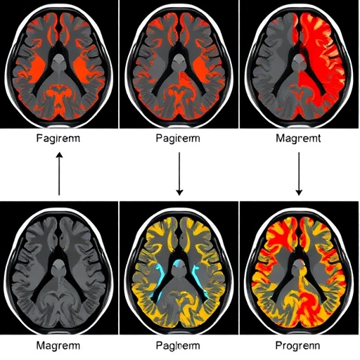
When a person arrives at the emergency department with the signature signs of an acute ischemic stroke, every minute matters. Each hour of delayed treatment costs the brain an estimated 1.9 million neurons, and the imaging decisions made in those first moments—whether the infarct is large, whether salvageable tissue remains, whether the patient should be rushed to the angiography suite for mechanical thrombectomy—can determine whether they walk out of the hospital or spend the rest of their life disabled. Yet the task at the heart of those decisions, tracing the exact boundaries of the dead and dying tissue on a CT or MRI scan, has long been a bottleneck: manual segmentation is slow, subjective, and notoriously variable from one radiologist to the next. A major new systematic review and meta-analysis published in BioMedical Engineering OnLine now offers the most comprehensive quantitative picture to date of how well artificial intelligence can take over that job—and the answer, on paper, is remarkably well.
The review, led by an international team of researchers from institutions spanning Iran, the United States, and Turkey, screened 4,755 records and ultimately included 101 studies that applied machine learning to the pixel- or voxel-level delineation of acute ischemic stroke lesions on neuroimaging. Adhering to PRISMA 2020 guidelines and registered prospectively, the analysis pooled performance metrics using random-effects models with inverse-variance weighting and logit transformation to stabilize the variance of bounded metrics. The headline numbers are striking: a pooled Dice coefficient of 0.84 with essentially zero between-study heterogeneity (I² = 0%), a pooled area under the receiver operating curve of 0.91, an accuracy of 0.89, a sensitivity of 0.85, and a specificity of 0.93. In practical terms, the best-performing machine learning systems overlap with expert-drawn lesion masks roughly as consistently as experts overlap with each other—something no prior synthesis has demonstrated with this level of statistical rigor.
The architecture landscape behind those numbers tells its own story. Deep learning approaches, overwhelmingly dominated by U-Net and its many descendants, accounted for 78 percent of the included studies, while classical methods such as support vector machines and random forests made up the remaining 22 percent. Within the deep learning camp, the review cataloged an impressive taxonomy of variants: conventional 2D U-Nets, 3D U-Nets, residual and attention-guided U-Nets, DenseNet- and ResNet-enhanced hybrids, cascaded pipelines, and modality-fusion frameworks. Each of these designs, the authors note, was engineered to attack a specific recurring challenge in stroke imaging—small lesion size, severe class imbalance between infarcted and healthy tissue, the notoriously faint contrast of early infarcts on non-contrast CT, and the need to fuse complementary MRI sequences or CT perfusion maps into a single decision. Two-dimensional implementations outpaced three-dimensional ones by more than two to one (68.6 percent versus 31.4 percent), and transformer-based and hybrid CNN-transformer models, despite generating considerable enthusiasm elsewhere in medical imaging, remained rare and under-validated in this domain.
Imaging modality emerged as one of the clearest determinants of success. In the subgroup meta-analysis, models built on diffusion-weighted imaging combined with apparent diffusion coefficient maps—the MRI sequences on which acute infarcts appear as bright, well-demarcated regions—achieved the highest pooled Dice scores, followed by MRI-only and multimodal MRI approaches. At the other end of the spectrum, studies relying on non-contrast CT alone or on CT angiography and CT perfusion data showed consistently lower pooled performance. The reason, the authors argue, is less a failure of the modality itself than a difference in what the models are being asked to do. Early ischemic changes on plain CT can be nearly invisible, while CT perfusion segmentation frequently amounts to a cross-modality prediction problem: the model must anticipate a lesion defined on follow-up or diffusion MRI from dynamic perfusion maps such as cerebral blood flow, cerebral blood volume, mean transit time, and Tmax. In benchmark challenges like ISLES 2018, the reference standard was the diffusion-defined infarct core rather than anything directly visible on the perfusion scan—a biologically evolving target rather than a static anatomical boundary. Lower scores there, the review cautions, should not be read as evidence that CT perfusion is inherently unsuited to machine learning.
Perhaps the most counterintuitive finding concerns data. A persistent assumption in machine learning is that more training data yields better models, but the meta-regressions here found no significant association between total image count and Dice performance. Most studies relied on fewer than 1,000 images, with only a handful exceeding 10,000, yet dataset size alone failed to explain the variation in results. Lesion volume fared no better: the correlation between study-level mean lesion volume and segmentation accuracy was weak and statistically indistinguishable from zero (r = −0.04, p = 0.80), though the authors stress this is an ecological, study-level association rather than proof that lesion size is irrelevant for individual patients. Meanwhile, a weak positive trend emerged between clinical severity, measured on the modified Rankin Scale, and segmentation performance (coefficient 0.022, p = 0.093)—an intriguing hint that larger, more conspicuous infarcts in severely affected patients may actually be easier for algorithms to find. Demographic continuity between training and test sets was solid: a Bland–Altman analysis of mean ages showed a negligible bias of −0.76 years, and training and test cohorts tracked each other closely (R² ≈ 0.85).
Beneath the headline metrics, the correlation analysis uncovered a subtler and clinically consequential trade-off. Dice coefficient, F1 score, and recall were tightly intercorrelated, as expected from their shared mathematical structure (Spearman ρ values of 0.95 and 0.82). Specificity, however, correlated weakly—and in some cases negatively—with these sensitivity-oriented metrics. In other words, models tuned aggressively to minimize false positives tend to miss subtle infarcts, a dangerous failure mode in wake-up strokes and posterior circulation events where lesions are small and easily overlooked. The authors argue that this trade-off undermines the common practice of judging stroke-segmentation models on a single headline number and calls for multi-dimensional evaluation frameworks that explicitly report the sensitivity-specificity balance alongside Dice and AUC.
The quality assessment, conducted with a modified QUADAS-2 tool, tempers the enthusiasm. While 65 of the 101 studies carried low risk of bias in patient selection and 72 in index test application, the reference standard and flow-and-timing domains were riddled with uncertainty: 22 studies were rated high risk and 19 unclear on the reference standard, and 25 high and 21 unclear on flow and timing. Inconsistently reported implementation details—loss functions, optimizers, learning-rate schedules, data augmentation, and the frequent conflation of patient counts with slice or augmented-image counts—further limited the meta-analysis and, more importantly, limit reproducibility across the field. The funnel plot for Dice scores showed no strong asymmetry, offering some reassurance against publication bias, though formal testing was not feasible.
The clinical implication of all this is a field that is algorithmically mature but translationally stalled. Despite pooled performance that would satisfy most regulatory benchmarks, machine learning segmentation is not yet routinely embedded in stroke triage workflows, automated ASPECTS scoring platforms, or thrombectomy eligibility algorithms. The authors point to the absence of prospective, multicenter validation across diverse scanners and populations, the lack of real-time processing benchmarks, and the scarcity of studies reporting clinically meaningful endpoints such as time saved, diagnostic confidence gained, or prognostic value added. Most models were trained and tested on retrospective, single-center datasets, and very few integrated clinical metadata such as NIHSS scores or comorbidities alongside the imaging. The review’s comparison with prior work underscores how far segmentation has come relative to prediction: an earlier meta-analysis of infarct prediction models reported a pooled Dice of just 0.50, whereas this segmentation-focused synthesis found 0.84—evidence, the authors suggest, that segmentation is the relatively mature corner of AI in stroke imaging, even as predictive modeling of tissue outcome remains far harder.
The path forward, the researchers argue, runs on two parallel tracks. For MRI-based models that already perform strongly, the priority shifts from architectural novelty to external validation, harmonized reporting standards, public benchmarking, workflow integration, and interpretable outputs—possibly accelerated through federated learning that allows models to train across institutions without moving patient data. For CT-based segmentation, where early lesions are subtle and perfusion targets evolve over hours, genuine technical innovation is still needed. Either way, the review concludes, the convergence of high pooled accuracy, zero heterogeneity in Dice scores, and a clearly mapped performance landscape now gives clinicians, developers, and regulators a common yardstick. The algorithms, it appears, are ready to read the scans. What remains is the harder work of proving they can do it—prospectively, in real emergency rooms, for every kind of stroke, on every scanner, in time to matter.
Subject of Research: Machine learning-based segmentation of acute ischemic stroke lesions on CT and MRI neuroimaging
Subject of Research: Medicine
Article Title: Systematic Review Weighs Machine Learning for Acute Ischemic Stroke Segmentation
Article References: Salehi, S., Birzhandi, S., Torbati Aghdam, Z., Amirvala, R., Dasoomi, H., M.Yaghi, Z., Soleimani Meigoli, M. S., Moghari Hesari, Z., Taghipoor, K., saffarian, K., Shahba, M., Ghaedamini, A., Ghazanafar Ahari, S., Arab Bafrani, M., Akhbari, M., Babapour, A., Rigi, A., Peiravi, S., & Asadi Anar, M. (2026). Machine learning models of segmentation in acute ischemic stroke: a systematic review and meta-analysis. BioMedical Engineering OnLine, 25(1), Article 101. https://doi.org/10.1186/s12938-026-01590-x
Image Credits: AI Generated
DOI: 10.1186/s12938-026-01590-x
Keywords: acute ischemic stroke segmentation, AI in emergency stroke management, AI-based brain imaging analysis, automated stroke lesion segmentation, brain tissue salvage prediction, CT and MRI stroke imaging, deep learning for ischemic stroke, machine learning in stroke diagnosis, medical image analysis systematic review, neural networks for stroke detection, neuroimaging segmentation accuracy, stroke treatment decision support
Cite Scienmag News
APA MLA Chicago
Cassandra Pierce. (September 8, 2026). Systematic Review Weighs Machine Learning for Acute Ischemic Stroke Segmentation. Scienmag. https://scienmag.com/systematic-review-weighs-machine-learning-for-acute-ischemic-stroke-segmentation/
Cassandra Pierce. “Systematic Review Weighs Machine Learning for Acute Ischemic Stroke Segmentation.” Scienmag, 8 September 2026, https://scienmag.com/systematic-review-weighs-machine-learning-for-acute-ischemic-stroke-segmentation/. Accessed 8 September 2026.
Cassandra Pierce. “Systematic Review Weighs Machine Learning for Acute Ischemic Stroke Segmentation.” Scienmag. September 8, 2026. https://scienmag.com/systematic-review-weighs-machine-learning-for-acute-ischemic-stroke-segmentation/
Copy citation Download RIS
Tags: acute ischemic stroke segmentationAI for medical image analysisAI in emergency stroke managementAI-based brain imaging analysisAI-driven neuroimaging decision supportautomated brain tissue segmentationautomated stroke lesion segmentationbrain tissue salvage predictionCT and MRI stroke imagingdeep learning for ischemic strokemachine learning in stroke diagnosismachine learning in stroke imagingmedical image analysis systematic reviewmedical image segmentation accuracyMRI and CT stroke segmentationneural networks for ischemic strokeneural networks for stroke detectionneuroimaging segmentation accuracyrapid stroke diagnosis with AIstroke lesion detection algorithmsstroke treatment decision supportstroke treatment planning with machine learningsystematic review of AI in stroke care