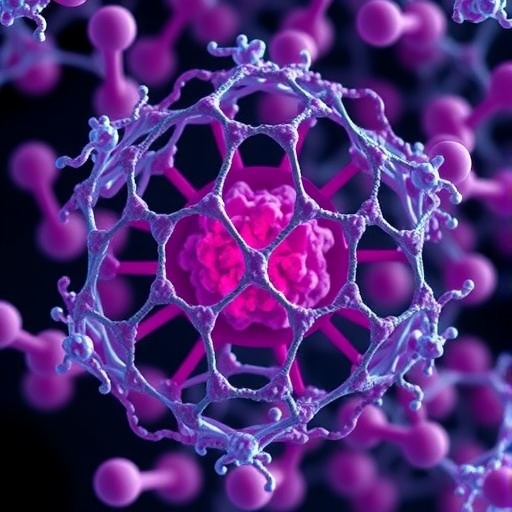
In a discovery that is reshaping how scientists think about the biology of human reproduction, researchers have unveiled the molecular architecture and three-dimensional spatial organization of proteasomes inside the nucleus of human sperm cells. The findings, published in Nature Structural & Molecular Biology, reveal that these protein-degrading molecular machines, long studied primarily for their housekeeping roles in the cytoplasm of dividing cells, occupy a precisely organized position within the densely packed paternal genome, where they appear to perform specialized functions that are essential for the earliest moments of embryonic development. The work, led by Paula Kolata, Álvaro dos Santos, Oliver Knowles, and colleagues, provides the most detailed structural picture to date of proteasomes in a cellular context that has remained remarkably resistant to conventional forms of biological analysis.
The human sperm cell is one of the most extraordinary cellular structures in biology. During spermatogenesis, the male germ cell strips away nearly all of its cytoplasm, condenses its genome to an extraordinary degree, and replaces the canonical histone-based chromatin of ordinary cells with protamines, small, highly basic proteins that allow DNA to be packed into a volume far smaller than that of a mitotic chromosome. This compaction is so extreme that the sperm head represents one of the most tightly organized packages of genetic material found in nature. While this architecture serves the practical purpose of delivering an intact paternal genome to the egg, it has long posed a formidable technical challenge: many of the standard tools of cell biology, from live-cell imaging to biochemical fractionation, function poorly or not at all in this context. As a result, the molecular inventory of the sperm nucleus, and especially the identity and arrangement of non-histone protein complexes within it, has remained only partially mapped.
Proteasomes themselves are among the most intensively studied molecular machines in biology. The 26S proteasome, the principal engine of intracellular protein degradation, consists of a barrel-shaped 20S catalytic core capped at one or both ends by 19S regulatory particles that recognize ubiquitin-tagged substrates, unfold them, and thread them into the proteolytic chamber for destruction. In somatic cells, proteasomes are distributed throughout the cytoplasm and nucleus, where they control protein turnover, regulate the cell cycle, and participate in transcriptional regulation and DNA repair. That proteasomes might exist inside the sperm nucleus was not entirely unexpected; earlier biochemical and immunological studies had detected proteasomal components in mature sperm from a variety of species. What had been missing was a coherent structural and spatial picture: were these simply residual contaminants from the discarded cytoplasm, or were they deliberately retained, correctly assembled, and spatially organized machines with a defined nuclear job?
To answer that question, the research team combined complementary approaches spanning structural biology, advanced microscopy, and spatial mapping techniques. Cryo-electron microscopy allowed the investigators to determine the molecular architecture of the proteasomal complexes associated with the sperm nucleus at near-atomic resolution, confirming that the structures present were not degraded remnants or misassembled fragments but intact, mature proteasomes with recognizable 20S cores and regulatory caps. In parallel, super-resolution and expansion microscopy approaches enabled the team to localize these complexes within the three-dimensional geometry of the sperm head, revealing that proteasomes are not randomly scattered through the nuclear volume but instead occupy reproducible, spatially patterned positions relative to the compacted chromatin. This spatial organization, the authors report, is consistent with a functional rather than incidental presence, suggesting that the paternal genome is delivered to the egg accompanied by an embedded, architecturally organized protein-degradation apparatus.
The significance of this spatial patterning becomes clearer when one considers what must happen in the minutes and hours following fertilization. When a sperm enters the egg, its densely protamine-packed chromatin must be rapidly remodeled: protamines are removed, histones are deposited, the paternal pronucleus forms, and the paternal genome must become competent for DNA replication and transcription. This sweeping decondensation is not a passive unraveling. It depends on the controlled modification and degradation of specific sperm-associated proteins, including protamines themselves in many species, and it has long been suspected that proteasomal activity contributes to this transition. Evidence from several mammalian systems has shown that inhibiting proteasome function can impair sperm capacitation, the acrosome reaction, and the ability of sperm to fertilize the egg. The new structural data provide a physical basis for these functional observations, showing that the machinery required for targeted protein destruction is not merely present in sperm but is positioned in the nucleus where the substrates of fertilization-linked remodeling reside.
One of the most provocative implications of the study concerns the origin and persistence of nuclear proteasomes through spermatogenesis. During the final stages of sperm differentiation, transcription ceases, the nuclear envelope is reorganized, and most nuclear functions are shut down. Yet the proteasomes identified in mature sperm appear fully assembled and structurally intact, raising the question of when they are imported or retained and how they remain stable through the dramatic nuclear remodeling that characterizes spermiogenesis. The authors’ architectural analysis suggests that these complexes may be loaded into the nucleus during earlier stages of sperm differentiation and preserved in a pre-armed state, ready to execute their degradative program at the moment of fertilization. This would represent a striking example of anticipatory cellular engineering, in which a cell packages not only its genome but also the molecular tools needed to unpack it after it reaches its destination.
The structural work also carries implications for how proteasomes interact with their substrates in a chromatin-dense environment. In somatic nuclei, proteasome access to chromatin is governed by a dynamic balance between freely diffusing and chromatin-bound populations. The sperm nucleus represents an extreme case of chromatin crowding, in which the available interchromosomal space is minimal and diffusion is severely constrained. The spatial map produced by the team indicates that nuclear proteasomes in sperm occupy defined microdomains, potentially corresponding to regions of the genome that retain histones or other non-protamine proteins susceptible to ubiquitin-mediated degradation. If confirmed, this organization would imply that the paternal genome is not uniformly packaged but instead contains differentiated zones, some destined for rapid proteasome-dependent remodeling and others for alternative histone-removal pathways. Such zonation would add a previously unrecognized layer of information to the sperm epigenome, one written not in chemical marks on DNA but in the physical placement of protein-processing machinery.
The findings may also open new avenues in reproductive medicine and fertility research. A substantial fraction of male infertility cases remain unexplained even after standard semen analysis, suggesting that defects in the fine-grained molecular content of sperm, beyond sperm count and motility, contribute to reproductive failure. Because proteasomal function has been implicated in sperm-zona pellucida interactions and in post-fertilization paternal genome remodeling, variations in the abundance, assembly state, or spatial deployment of nuclear proteasomes could plausibly affect embryonic development before the paternal genome is even activated. The ability to characterize proteasome architecture in human sperm, as demonstrated in this study, provides a template for investigating whether such molecular signatures correlate with fertilization success, embryo quality, or recurrent pregnancy loss. It also raises the possibility of pharmacological interventions, since the proteasome is a well-established drug target in oncology and its inhibitors and modulators are extensively characterized.
Beyond reproductive biology, the study speaks to a broader conceptual shift in how the field understands the sperm cell. For much of the twentieth century, the sperm was viewed as a streamlined delivery vehicle, its biology largely reduced to locomotion and membrane fusion. Over the past two decades, that view has given way to a far richer picture in which sperm carry retained centrosomal proteins, small RNAs, DNA methylation patterns, histone modifications, and now organized nuclear protein-degradation machinery, all of which may influence the developmental program of the embryo. The demonstration that a major molecular machine of the protein-quality-control system is architecturally integrated into the sperm nucleus underscores that paternal contributions to the zygote extend well beyond the raw sequence of DNA. The paternal genome, in effect, arrives with its own unpacking apparatus.
Technically, the study also demonstrates the growing power of structural and imaging methods to interrogate biological systems that were previously considered inaccessible. The sperm nucleus is small, optically dense, biochemically intractable, and cannot be genetically manipulated in mature form, which has historically limited progress. By extracting architectural information through cryo-electron microscopy and coupling it with spatially resolved localization methods, the team has shown that a detailed molecular census of this unusual nuclear environment is achievable. The approach is likely to be applied to other retained nuclear complexes in sperm, and possibly to other highly condensed chromatin systems, such as the nuclei of certain protozoa or the mitotic chromosomes of early embryos, where the interplay between compaction and protein turnover remains poorly understood.
Looking forward, the authors and others in the field will aim to identify the full set of substrates targeted by sperm nuclear proteasomes, to determine how proteasome positioning is established during spermatogenesis, and to test directly whether disruption of nuclear proteasome organization produces measurable fertility defects. Each of these questions builds on the foundation laid by the current work. What is already clear is that the human sperm nucleus, far from being an inert container of DNA, is a precisely engineered structure in which genomes and protein-degradation machines are co-organized with apparent purpose. That realization reframes fertilization itself: the encounter between sperm and egg is not simply the meeting of two genomes but the coordinated activation of two elaborate molecular programs, one of which, it now appears, arrives pre-installed inside the paternal nucleus, ready to begin dismantling and rebuilding the paternal chromatin at the very moment a new life begins.
Subject of Research: Molecular architecture and spatial organization of proteasomes within the human sperm nucleus
Subject of Research: Biology
Article Title: Molecular architecture and spatial organization of proteasomes in the human sperm nucleus
Article References: Kolata, P., dos Santos, Á., Knowles, O., Dendooven, T., & Allegretti, M. (2026). Molecular architecture and spatial organization of proteasomes in the human sperm nucleus. Nature Structural & Molecular Biology. https://doi.org/10.1038/s41594-026-01870-z
Image Credits: AI Generated
DOI: 10.1038/s41594-026-01870-z
Keywords: proteasome, human sperm nucleus, protamine, chromatin remodeling, fertilization, paternal genome, cryo-electron microscopy, spermatogenesis, male fertility, protein degradation, 26S proteasome, sperm epigenome
Cite Scienmag News
APA MLA Chicago
Drew Townsend. (September 7, 2026). Human sperm nucleus proteasomes revealed through molecular architecture and spatial organization. Scienmag. https://scienmag.com/human-sperm-nucleus-proteasomes-revealed-through-molecular-architecture-and-spatial-organization/
Drew Townsend. “Human sperm nucleus proteasomes revealed through molecular architecture and spatial organization.” Scienmag, 7 September 2026, https://scienmag.com/human-sperm-nucleus-proteasomes-revealed-through-molecular-architecture-and-spatial-organization/. Accessed 7 September 2026.
Drew Townsend. “Human sperm nucleus proteasomes revealed through molecular architecture and spatial organization.” Scienmag. September 7, 2026. https://scienmag.com/human-sperm-nucleus-proteasomes-revealed-through-molecular-architecture-and-spatial-organization/
Copy citation Download RIS
Tags: 3D spatial organization of proteasomesadvanced imaging of proteasadvanced imaging of proteasomes in spermcellular organization of proteashuman sperm cell proteasome architecturehuman sperm nucleus proteasomesmolecular architecture of proteasomes in spermmolecular structure of sperm cell proteasomesproteasome distribution during spermatogenesisproteasome function in sperm chromatin remodelingproteasome functions during spermatogenesisproteasome localization in paternal genomeproteasome localization within densely packed paternal genomeproteasomes in sperm genome packagingproteasomes role in human reproductionprotein degradation machinery in human spermrole of proteasomes in human embryonic developmentspecialized functions of proteasomes in embryonic developmentspecialized functions of proteasomes in spermsperm nuclear proteasomesstructural analysis of proteasomes in reproductive cellsstructural analysis of sperm cell proteasomesthree-dimensional spatial organization of proteasomes in sperm nuclei