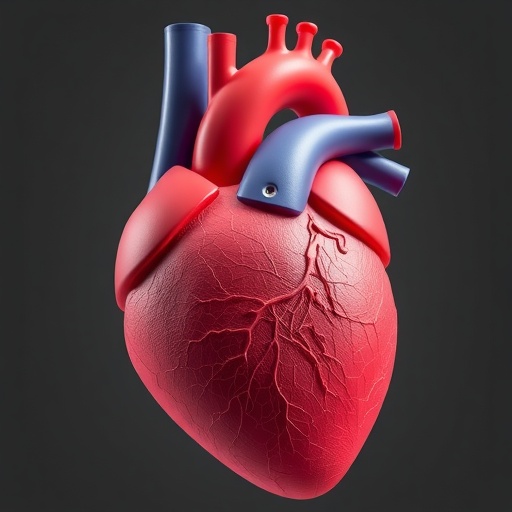
Heart surgeons in Poland have shown how a digital twin of a beating heart can do far more than guide a delicate operation—it can also prevent a patient from being sent back to the operating table unnecessarily. In a case report published in the journal 3D Printing in Medicine, a team at the Medical University of Białystok describes how three-dimensional visualization, modeling, and printing shaped both the surgical treatment and the rocky postoperative course of a 49-year-old man with hypertrophic obstructive cardiomyopathy, a thickening of the heart muscle that blocks blood from leaving the heart’s main pumping chamber.
Hypertrophic obstructive cardiomyopathy, often abbreviated HOCM, is among the most common inherited cardiac conditions, affecting roughly one in every 500 people. In many patients, the muscular wall that separates the two ventricles—the interventricular septum—becomes abnormally thick, narrowing the left ventricular outflow tract, the passageway through which oxygenated blood exits the heart toward the aorta. During each heartbeat, the obstruction can be worsened by a phenomenon known as systolic anterior motion, in which the mitral valve leaflet is dragged into the narrowed tract, further obstructing flow and often causing leakage of the valve. When medications fail, surgeons can remove part of the thickened septum in an operation called septal myectomy—a procedure that demands precise judgment, since cutting too little leaves the obstruction behind and cutting too much risks damaging the heart’s electrical and structural machinery.
Traditionally, surgeons plan this operation using two-dimensional echocardiographic images and angiographic slices, mentally reconstructing the complex three-dimensional geometry of the ventricle. The Białystok team took a different approach. They began with cardiac computed tomography angiography, a scan that captures the patient’s heart in exquisite anatomical detail, and then segmented the imaging data to build a virtual three-dimensional model of the left ventricular cavity and the obstructed outflow tract. On this digital replica, the surgeons performed a virtual myectomy, rehearsing exactly which portion of the septum to resect before ever entering the operating room. They also printed patient-specific physical models of the heart, which were sterilized in an unusual way—using hydrogen peroxide plasma, a low-temperature sterilization process that can render delicate printed materials safe for the sterile surgical field without deforming them.
The printed models accompanied the surgical team into the operating room, where they served as tactile anatomical guides during the extended septal myectomy. Rather than relying solely on the surgeon’s mental image of the anatomy, the team could hold the patient’s actual ventricular geometry in their hands, correlating what they felt and saw with the digital plan. Intraoperative echocardiography performed at the end of the operation delivered exactly the result surgeons hope for: the systolic anterior motion of the mitral valve had resolved, and the pressure gradient across the left ventricular outflow tract—the hemodynamic measure of the obstruction—had dropped to a mere 6 mmHg, well within the range considered a successful relief of obstruction.
What happened next is what makes this case report stand out. On the third day after surgery, the patient suddenly became short of breath again. Echocardiography revealed severe recurrence of the outflow tract obstruction, with a peak gradient of 73 mmHg—higher than many patients experience before surgery—and the return of systolic anterior motion of the mitral valve. To any surgical team, this is a moment of high tension: is there residual muscle left behind that must be removed in a second operation, or is something else at work?
Rather than rushing back to surgery, the team repeated the cardiac computed tomography angiography and rebuilt their three-dimensional models. The new digital reconstruction showed something decisive: there was no residual anatomic substrate for obstruction. The basal septum measured a maximum of 15 mm in thickness, a value that, after adequate myectomy, leaves no structural target for further resection. The anatomy was fixed; the physiology was not. The 3D evidence pointed instead to a dynamic mechanism—a hyperdynamic, vigorously contracting left ventricle, provoked by relative intravascular hypovolemia (too little blood volume in the vessels), tachycardia (an abnormally fast heart rate), and the vasodilating drugs the patient had been receiving. In this state, even a normally resected outflow tract can become obstructed, because the chamber empties too forcefully and too quickly.
Armed with that understanding, the clinicians shifted strategy entirely away from the scalpel. They treated the patient with hemodynamically targeted conservative management: augmenting preload by giving intravenous fluid to fill the ventricle more completely, discontinuing vasodilatory therapy that was lowering vascular resistance and fueling hyperdynamic contraction, and intensifying heart-rate control to slow the ventricle down. Each intervention directly countered one element of the dynamic obstruction identified through imaging. The response was dramatic and measurable. By postoperative day 7, the outflow tract gradient had normalized to 12 mmHg, the systolic anterior motion had resolved again, and the patient’s functional capacity improved steadily.
The clinical lesson, the authors argue, is that 3D modeling should not be viewed as a purely preoperative luxury. When repeated after surgery, the same segmentation and reconstruction pipeline can distinguish a structural problem—which would warrant reintervention—from a physiological problem, which calls for medical management. That distinction carries real weight, because early postoperative gradients in these patients can look alarming, and a premature second operation would expose the patient to serious risk while addressing, in this case, a problem that fluids and beta-blockers could resolve. The case also illustrates the complementary value of the physical prints, processed with hydrogen peroxide plasma sterilization, in confirming resection adequacy during the operation itself.
The team behind the report spans cardiosurgery, radiology, cardiology, and even a students’ scientific club at the Medical University of Białystok, led by corresponding author Miłosz Nesterowicz and first author Uladzimir Andrushchuk. Their case adds to a growing body of evidence that patient-specific 3D printing, once considered an experimental novelty, is maturing into practical clinical infrastructure—useful for rehearsal, for intraoperative navigation, and now for postoperative diagnostic reasoning. For a condition in which the line between a successful operation and a catastrophic complication can hinge on millimeters of muscle and the dynamics of a single heartbeat, having the heart in three dimensions, both on screen and in hand, may prove to be one of the most consequential tools in the cardiac surgeon’s kit.
The patient, whose written informed consent made publication possible, ultimately avoided reintervention and recovered with restored outflow tract gradients and improved exercise capacity. As the technology becomes more accessible—segmentation software, printing materials, and sterilization protocols are all steadily improving—cases like this one suggest that the question is no longer whether 3D modeling belongs in cardiac surgery, but how routinely it should be woven into every stage of care, from the first scan to the last follow-up echocardiogram.
Subject of Research: Use of 3D visualization, modeling, and printing for surgical planning and postoperative decision-making in hypertrophic obstructive cardiomyopathy
Subject of Research: Medicine
Article Title: 3D visualization and modeling in the surgical treatment and postoperative management of hypertrophic obstructive cardiomyopathy: a case report
Article References: Andrushchuk, U., Garbowska, M., Chrostowski, T., Andrushchuk, D., Dzieżyk, W., Sulima, D., Litvinenko, A., & Nesterowicz, M. (2026). 3D visualization and modeling in the surgical treatment and postoperative management of hypertrophic obstructive cardiomyopathy: a case report. 3D Printing in Medicine. https://doi.org/10.1186/s41205-026-00348-4
Image Credits: AI Generated
DOI: 10.1186/s41205-026-00348-4
Keywords: 3D visualization, 3D modeling, 3D printing, cardiac computed tomography angiography, hypertrophic obstructive cardiomyopathy, septal myectomy, dynamic left ventricular outflow tract obstruction, case report
Cite Scienmag News
APA MLA Chicago
Ophelia Keating. (September 4, 2026). 3D modeling guides surgery and care in hypertrophic obstructive cardiomyopathy case. Scienmag. https://scienmag.com/3d-modeling-guides-surgery-and-care-in-hypertrophic-obstructive-cardiomyopathy-case/
Ophelia Keating. “3D modeling guides surgery and care in hypertrophic obstructive cardiomyopathy case.” Scienmag, 4 September 2026, https://scienmag.com/3d-modeling-guides-surgery-and-care-in-hypertrophic-obstructive-cardiomyopathy-case/. Accessed 4 September 2026.
Ophelia Keating. “3D modeling guides surgery and care in hypertrophic obstructive cardiomyopathy case.” Scienmag. September 4, 2026. https://scienmag.com/3d-modeling-guides-surgery-and-care-in-hypertrophic-obstructive-cardiomyopathy-case/
Copy citation Download RIS
Tags: 3D heart modeling3D heart modeling for hypertrophic obstructive cardiomyopathy3D printing in cardiac surgical planning3D printing in medical surgery3D visualization for cardiac valve leaflet movement3D visualization in hypertrophic cardiomyopathy treatmentdigital twin in cardiac surgeryheart anatomy modelinghypertrophic obstructive cardiomyopathy treatmentintervention planning for septal hypertrophyinterventricular septum modelingleft ventricular outflow tract obstruction managementminimally invasive heart surgery guidanceminimally invasive HOCM surgical guidespersonalized cardiac surgical approachespost-operative care in cardiac surgerypostoperative care in hypertrophic cardiomyopathypreventing reoperation in HOCM patientsrole of 3D printing in cardiac diseasesurgical intervention for thickened heart musclesurgical planning for HOCMvisualization of hypertrophic cardiomyopathy