Cerebral amyloid angiopathy has long been defined by its most feared manifestation: bleeding into the brain. For decades, clinicians and researchers have treated this age-related disease of the brain’s small vessels as a fundamentally hemorrhagic disorder, in which amyloid protein deposits stiffen and fragilize vessel walls until they rupture, producing lobar intracerebral hemorrhage and microbleeds visible on MRI. But a new study from Peking Union Medical College Hospital, published in the Journal of Neurology, adds weight to an increasingly insistent counter-argument: the brain under CAA is also quietly ischemic, suffering small strokes that are sometimes dramatic and sometimes completely silent, and that these two faces of ischemia follow strikingly different rules.
The study, led by Yuhui Sha and Jun Ni, together with collaborators including Joanna M. Wardlaw of the University of Edinburgh, set out to systematically characterize symptomatic diffusion-weighted imaging lesions, known as sDWI lesions, and incidental DWI lesions, known as iDWI lesions, in patients with probable CAA. Diffusion-weighted imaging is an MRI sequence exquisitely sensitive to acute cellular injury: when blood flow to brain tissue fails and cells begin to swell with cytotoxic edema, water molecules lose their freedom to diffuse, and the affected region lights up brightly on the scan. In CAA, these bright spots have been the subject of a running debate. Autopsy work from previous groups, including studies by Ter Telgte and colleagues, has suggested that DWI-positive lesions in CAA correspond histologically to microinfarcts, confirming that they represent genuine ischemia rather than hemorrhage or artifact. Yet the clinical relevance, prevalence, and mechanisms of these lesions remained contested.
To resolve the question, the team drew on a prospective cohort of cerebral small vessel disease patients recruited at Peking Union Medical College Hospital between March 2017 and February 2026. Using the Boston criteria version 1.5, the standard clinicoradiological framework for diagnosing CAA without tissue biopsy, they enrolled 185 patients with probable CAA. Each participant underwent detailed baseline clinical assessment and multimodal MRI, including imaging markers of small vessel disease such as lacunes, cerebral microbleeds, and cortical superficial siderosis, the latter being a sign of chronic blood leaking over the surface of the brain. Ninety-nine of the patients returned for follow-up MRI, generating 163 scans in total, which allowed the researchers to estimate not only cross-sectional prevalence but also cumulative incidence over time.
The results were revealing in two directions. First, ischemia turned out to be anything but rare. Symptomatic DWI lesions were detected in 29 patients, while incidental DWI lesions, discovered on scans performed for other reasons, appeared in 49 patients, more than a quarter of the entire cohort. Kaplan–Meier analysis of the followed subgroup showed that over three years, the cumulative incidence of new symptomatic DWI lesions was 9.45 percent, with a 95 percent confidence interval of 0.67 to 17.46 percent, while the cumulative incidence of incidental lesions was considerably higher at 25.74 percent, with a confidence interval spanning 15.2 to 34.97 percent. In other words, roughly one in four CAA patients followed longitudinally was destined to develop a small ischemic lesion within three years, most of them without any clinical fanfare.
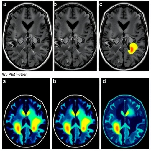
Second, and perhaps more striking, the two lesion types behaved like two different diseases. When the researchers constructed lesion probability maps to visualize topography, symptomatic DWI lesions clustered predominantly in deep brain regions, accounting for 72.7 percent of their locations. Incidental lesions, by contrast, were mainly found in cortical and juxtacortical regions, the ribbon of gray matter and the immediately underlying white matter, in 62.5 percent of cases. This anatomical split is not trivial. Deep and superficial cerebral territories are supplied by different vascular trees, with deep perforating arterioles and leptomeningeal cortical vessels subject to different hemodynamic stresses and pathological processes. A preferential distribution suggests a preferential mechanism.
That suspicion was confirmed by prospective Cox regression analysis, which adjusted for age and sex and, for the incidental lesions, also for traditional vascular risk factors and focal superficial siderosis. For symptomatic DWI lesions, the independent predictors were the number of traditional vascular risk factors, including hypertension, diabetes mellitus, coronary artery disease, and smoking, with a hazard ratio of 2.968 per additional risk factor, and the presence of lacunes, small old infarcts typical of hypertensive arteriopathy, with a hazard ratio of 1.187. Both reached conventional statistical significance, with P values of 0.013 and 0.016 respectively. This profile points toward the familiar machinery of atherosclerotic and hypertensive small vessel disease, superimposed on the amyloid-laden vasculature of CAA patients.
Incidental DWI lesions told a different story. Their independent predictors were lacunes, with a hazard ratio of 1.158, disseminated cortical superficial siderosis, with a hazard ratio of 2.994, and lobar cerebral microbleed grade, with a hazard ratio of 2.059. Notably, traditional vascular risk factors did not independently predict these silent lesions once adjustments were made. Disseminated siderosis and a heavy burden of lobar microbleeds are both hallmarks of advanced amyloid pathology, marking vessels that leak blood products into the subarachnoid space and brain parenchyma. Their association with cortical and juxtacortical ischemic lesions fits a mechanistic picture in which amyloid-laden leptomeningeal and cortical vessels, damaged by both amyloid deposition and hemosiderin-related injury, suffer episodic failure of perfusion, producing tiny cortical microinfarcts that the patient never notices.
The implications ripple outward in several directions. Clinically, the findings suggest that when a CAA patient presents with an acute neurological event, a DWI-positive lesion in deep structures is likely to be associated with conventional vascular risk factors and may warrant aggressive management of those factors. Conversely, incidental cortical lesions in a patient with disseminated siderosis may signal active amyloid-related vascular injury, which has been linked in prior work to increased risks of both future hemorrhage and cognitive decline. Earlier studies have shown that silent ischemic infarcts are associated with hemorrhage burden in CAA, and that DWI lesions after intracerebral hemorrhage predict recurrent stroke, so the new prospective risk factor data give clinicians a sharper tool for risk stratification.
Scientifically, the study reframes DWI-positive lesions as a dynamic window on small vessel disease activity. Traditional imaging markers such as microbleeds and siderosis are cumulative, recording injury that may have accumulated over years. DWI lesions, by contrast, are acute, appearing and resolving within days to weeks, and their signal characteristics evolve in a characteristic sequence as microinfarcts mature. Their detection rate in a longitudinal cohort therefore reflects ongoing disease activity, in the same way that incident troponin release reflects active myocardial injury. The authors propose that DWI-positive lesions could serve as a useful imaging marker of SVD activity and injury in future longitudinal studies and clinical trials of CAA. This matters because the CAA therapeutic pipeline is, for the first time, showing real signs of life. Immunotherapy trials with anti-amyloid antibodies such as ponezumab have been conducted, and a Phase 2 study of the RNA interference therapeutic ALN-APP, designed to reduce production of amyloid precursor protein, is currently recruiting patients with CAA. Trials like these need sensitive, quantifiable markers of target engagement and disease progression, and DWI lesion rates could fill that role.
There are, as always, caveats. The cohort of 185 patients, and the 99 with follow-up imaging, is modest by the standards of national registry studies, and the confidence interval around the three-year incidence of symptomatic lesions is wide, reflecting the relative rarity of these events. Probable CAA by Boston criteria is not histologically confirmed disease, although the criteria perform well in autopsy-validated settings. The observational design cannot definitively separate correlation from causation: lacunes may predict DWI lesions because they share upstream causes rather than because old infarcts directly provoke new ones. And MRI at typical clinical field strengths can miss very small cortical microinfarcts, meaning the true incidence of ischemia in CAA may be even higher than these figures suggest. Nevertheless, the consistency of the topographical and risk factor dissociation across analyses lends credibility to the central conclusion.
The broader message is a conceptual one. CAA, the study concludes, is not simply a hemorrhagic disease with an occasional ischemic afterthought. Cerebral ischemia is common, mechanistically patterned, and readable on standard clinical MRI. Symptomatic deep lesions track the burden of systemic vascular risk, while silent cortical lesions track the severity of amyloid vasculopathy itself, a dissociation that echoes recent work in CAA and CADASIL cohorts showing high prevalences of incidental DWI lesions with distinct clinical associations. For a disease whose diagnosis has historically hinged on documenting bleeding, the growing recognition that acute ischemic lesions can be counted, mapped, and followed over time offers both a warning and an opportunity: the amyloid-diseased vessel fails in more ways than one, and each failure mode now has its own signature that clinicians and trialists can learn to read.
Subject of Research: Prevalence, distribution, and risk factors of symptomatic and incidental diffusion-weighted imaging lesions indicating cerebral ischemia in cerebral amyloid angiopathy
Subject of Research: Medicine
Article Title: Distinct characteristics and risk factors of symptomatic and incidental DWI lesions in cerebral amyloid angiopathy
Article References: Sha, Y., Wu, J., Zhou, Y., Liu, Z., Han, F., Yao, M., Zhou, L., Zhu, Y., Wardlaw, J. M., & Ni, J. (2026). Distinct characteristics and risk factors of symptomatic and incidental DWI lesions in cerebral amyloid angiopathy. Journal of Neurology, 273(9), Article 559. https://doi.org/10.1007/s00415-026-14107-2
Image Credits: AI Generated
DOI: 10.1007/s00415-026-14107-2
Keywords: cerebral amyloid angiopathy, diffusion-weighted imaging, symptomatic DWI lesions, incidental DWI lesions, cerebral small vessel disease, cortical superficial siderosis, cerebral microbleeds, lacunes, microinfarcts, risk factors, vascular risk factors, longitudinal MRI
Cite Scienmag News
APA
MLA
Chicago
Cassandra Pierce. (September 4, 2026). Symptomatic, Incidental DWI Lesions Show Distinct Risk Factors in Cerebral Amyloid Angiopathy. Scienmag. https://scienmag.com/symptomatic-incidental-dwi-lesions-show-distinct-risk-factors-in-cerebral-amyloid-angiopathy/
Cassandra Pierce. “Symptomatic, Incidental DWI Lesions Show Distinct Risk Factors in Cerebral Amyloid Angiopathy.” Scienmag, 4 September 2026, https://scienmag.com/symptomatic-incidental-dwi-lesions-show-distinct-risk-factors-in-cerebral-amyloid-angiopathy/. Accessed 4 September 2026.
Cassandra Pierce. “Symptomatic, Incidental DWI Lesions Show Distinct Risk Factors in Cerebral Amyloid Angiopathy.” Scienmag. September 4, 2026. https://scienmag.com/symptomatic-incidental-dwi-lesions-show-distinct-risk-factors-in-cerebral-amyloid-angiopathy/
Copy citation
Download RIS
Tags: brain hemorrhage risk factorsCerebral amyloid angiopathycerebral microinfarctscerebrovascular disease mechanismsDWI lesionsincidental DWIincidental DWI lesionsischemic brain injuryischemic strokemicrobleedsmicrobleeds and hemorrhageMRI imaging in CAAMRI in neurovascular diseasesneuroimaging biomarkersneurovascular imagingrisk factors in CAAsilent cerebral ischemiasilent strokessmall vessel diseasesymptomatic diffusion-weighted imagingsymptomatic DWI