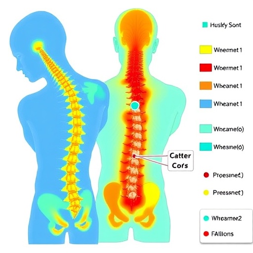
When a severe accident crushes a human spinal cord, the most dangerous hours unfold silently. Surgeons can realign the spine, relieve compression, and stabilize the column with rods and screws, but the cord itself — sealed inside bone and tough dura mater — gives clinicians no routine readout of its condition. While neurointensivists routinely track pressure, oxygen, and metabolism inside an injured brain, the injured spinal cord has remained one of critical care medicine’s last unmonitored organs. A new study published in the journal Neurocritical Care on 14 August 2026 argues that this blindness is a choice, not a necessity. An international team reports the largest and most geographically diverse experience yet with monitoring probes placed directly into the fluid space at the site of injury: 109 adults treated in five intensive care units across Europe. The probes generated good-quality signals in the overwhelming majority of patients, remained safely in place for an average of more than five days, and produced no cases of spinal cord damage, meningitis, or bleeding. The work, coordinated through the European SOPRANI network with co-senior authors Marios C. Papadopoulos and Samira Saadoun of City St George’s, University of London, provides the strongest evidence yet that direct monitoring of the injured human cord is feasible, informative, and acceptably safe.
The clinical problem is rooted in simple mechanics. After traumatic impact, the spinal cord swells with edema and hemorrhage, yet it is confined within the rigid vertebral canal and an inelastic dural sac — a closed compartment with almost no room to spare. As pressure within the injured segment rises, it squeezes the small vessels supplying the cord from the outside, and blood flow falls. Starved of oxygen, neural tissue slides into a cascade of secondary injury: excitotoxic neurotransmitter release, mitochondrial failure, free radical generation, and progressive cell death that can convert a partial injury into a complete, permanent one. Guidelines already recognize the stakes, recommending elevated blood pressure targets after acute injury, because systemic perfusion is currently the only lever clinicians can pull. But without knowing the pressure inside the injured cord itself, every patient is treated with the same blunt instrument: push the mean arterial pressure up and hope. Neurosurgeons in the field have long argued that this is equivalent to managing traumatic brain injury without measuring intracranial pressure — a practice medicine abandoned decades ago after it became clear how dangerous flying blind could be.
The technology described in the new paper adapts a method proven in the brain. During the initial decompression and stabilization operation, surgeons insert a slim pressure transducer — essentially the strain-gauge technology long used for intracranial pressure monitoring — through the dura so that the sensor sits intrathecally at the injured cord segment. The device reports intraspinal pressure, the pressure within the dural tube surrounding the cord, continuously and in real time. From it flows a second, more meaningful number: spinal cord perfusion pressure, calculated as mean arterial blood pressure minus intraspinal pressure, a direct analogue of the cerebral perfusion pressure formula that anchors head-injury intensive care. Perfusion pressure is the driving force pushing blood through the cord’s microcirculation; when it falls too low, ischemia follows no matter how reassuring the systemic vital signs appear. Because the probe samples many times per second, clinicians can also derive indices of spinal cord autoregulation — the injured vasculature’s capacity to hold blood flow constant as blood pressure drifts. In earlier single-center work, higher intraspinal pressures and lower perfusion pressures tracked with worse neurological recovery, evidence that the measurements capture meaningful physiology rather than electronic noise.
Pressure is only the first layer of information. In most patients the teams added a microdialysis catheter: a fine hollow fiber tipped with a semipermeable membrane, perfused at an ultra-slow rate with sterile artificial cerebrospinal fluid. Small molecules in the injured cord’s extracellular space — glucose, lactate, pyruvate, glycerol, and the excitatory amino acid glutamate — diffuse across the membrane into the perfusate, which is collected in microvials changed at the bedside and analyzed. The lactate-to-pyruvate ratio serves as a sensitive flag for cellular energy failure, rising when mitochondria cannot exploit oxygen and cells fall back on anaerobic metabolism; glycerol signals membrane breakdown and cell death; glutamate marks excitotoxic stress. Roughly two in five patients in the new series also carried a third probe measuring tissue oxygen tension directly within the injured cord, converting the abstract idea of cord oxygenation into a number on a monitor. Together, the three probes turn the injury site into something closer to an instrumented organ: pressure, perfusion, oxygen, and metabolism, all sampled continuously through the most dangerous days after trauma.
What separates the new report — first authored by Love C. Ilochonwu — from earlier single-center studies is its scale and diversity. The analysis pooled experience from intensive care units at Aarhus University Hospital in Denmark, University Hospitals Leuven in Belgium, University Medical Centre Ljubljana in Slovenia, St George’s Hospital in London, and Hospital Universitario 12 de Octubre in Madrid. The researchers distributed a standardized questionnaire to all participating sites and retrospectively extracted each patient’s course from the medical record. The 109 adults had sustained acute, severe traumatic spinal cord injuries, and the cohort was severely affected: 58.7% were grade A on the American Spinal Injury Association Impairment Scale, meaning complete injury with no motor or sensory function preserved below the lesion; 19.3% were grade B, with sensory but no motor function spared; and 22.0% were grade C. The probes, placed during the initial surgery, stayed in situ for 5.3 ± 0.2 days on average — a window spanning precisely the period when cord swelling peaks and hemodynamic instability is most dangerous, and long enough to generate dense physiological datasets for every patient.
The results answer the two questions skeptics ask first: do the probes work, and do they stay where surgeons put them? Every patient received an intraspinal pressure probe; 67.9% also received microdialysis, and 44.0% also received an oxygen probe. Signal quality was rated good most of the time in 89.0% of patients for pressure, 95.9% for microdialysis, and 79.2% for oxygen — a performance envelope comparable to established neuromonitoring in the brain. Placement was verified with computed tomography in 89.0% of patients, and the scans confirmed good probe position in every case but one. That record matters, because a probe that has migrated or malpositioned measures the wrong tissue or nothing at all, silently corrupting the numbers on which clinical decisions would rest. The oxygen sensors were the least consistent of the three technologies, but they still delivered good signals most of the time in nearly four of every five patients — a respectable figure for instruments that must function inside a swollen, moving, metabolically hostile environment for close to a week.
Safety was the study’s central question, and the findings are reassuring with important caveats. The most common complication was cerebrospinal fluid leak, seen in 20.8% of patients — an expected consequence of passing a probe through the dura. Most leaks were straightforward to manage: 81.8% required only bedside suturing, 9.1% needed temporary lumbar drainage of cerebrospinal fluid to offload pressure, and 9.1% required early removal of the probe. Pseudomeningocele, a pocket of cerebrospinal fluid collecting beneath the wound, developed in 10.4% of patients; 81.8% of these were asymptomatic and needed nothing more than observation, while 18.2% required surgery. Wound infections occurred in 4.7% of patients, resolving with antibiotics alone in 60.0% of cases and requiring surgical debridement in the remainder. Set against these manageable events is what the team never observed across the entire cohort: not one case of spinal cord damage from the probes themselves, not one meningitis, not one spinal hematoma. In the authors’ conclusion, the main risks of intrathecal monitoring are CSF leak, pseudomeningocele, and wound infection — recognizable, treatable complications — while the feared catastrophic events simply did not occur.
The most provocative material concerns what the probes reveal at the bedside. Although the signals in this series were collected primarily for research, the authors present examples of their potential clinical value: real-time visualization of spinal cord perfusion pressure, of cord autoregulation, and of tissue oxygenation and metabolism. In practice, that means an intensivist can watch whether raising blood pressure with fluids and vasopressors genuinely improves perfusion of the injured segment, or whether the cord’s vessels have lost the capacity to respond. It can expose the patient whose perfusion pressure looks adequate on paper while microdialysis shows a climbing lactate-to-pyruvate ratio — tissue starving despite acceptable numbers. It can reveal oxygen desaturation within the cord that no systemic monitor would detect. Earlier work by the London group found that intraspinal pressure and perfusion pressure measured in the first days after injury predicted neurological outcome, lending the signals prognostic weight as well as physiological meaning. The parallel with neurocritical care for the brain is hard to miss: intracranial pressure monitoring helped transform traumatic brain injury from intuitive management into protocolized, physiology-guided care, and spinal cord monitoring now stands roughly where brain monitoring stood a generation ago.
The authors are candid about limits. This was a retrospective case series without a control group; it establishes feasibility, signal quality, and safety, not benefit. Whether patients managed with monitoring-guided perfusion targets recover more function than those managed conventionally is precisely the question that prospective trials must now answer, and the multi-center infrastructure demonstrated here looks like scaffolding for exactly those studies. Retrospective data can also under-ascertain complications, although the thoroughness of the safety accounting and the consistency of results across five institutions, five surgical teams, and five intensive care cultures argue that the technique is robust beyond any single expert center. The research was supported by the Marie Skłodowska-Curie Actions SOPRANI network, Wings for Life, UK Research and Innovation, and the UK National Institute for Health and Care Research. For the hundreds of thousands of people who sustain traumatic spinal cord injuries worldwide each year, the immediate message is narrow but significant: the tools to see inside the injured cord exist, they work in ordinary intensive care units, and their risk profile is now quantified. The era of treating the injured spinal cord as a black box may finally be closing.
Subject of Research: Safety, placement accuracy, signal quality, and clinical utility of intrathecal monitoring probes — measuring intraspinal pressure, spinal cord perfusion pressure, tissue oxygen, and metabolism — placed at the injury site in 109 patients with acute, severe traumatic spinal cord injury across five European intensive care units.
Subject of Research: Medicine
Article Title: Monitoring from the Injury Site After Spinal Cord Injury: Case Series of 109 Patients from Five Intensive Care Units
Article References: Ilochonwu, L. C., Asif, H., Kopač, C., Jug, M., Depreitere, B., Sansinenea, I. P., Gómez-Abascal, A. L., Thygesen, M. M., Rasmussen, M. M., Bosetta, E., Zoumprouli, A., Papadopoulos, M. C., Saadoun, S., The SOPRANI Collaborators, Urban, A., Baud, E., Moberg, D., Meyfroidt, G., Depreitere, B., … Rehber, C. (2026). Monitoring from the Injury Site After Spinal Cord Injury: Case Series of 109 Patients from Five Intensive Care Units. Neurocritical Care. https://doi.org/10.1007/s12028-026-02629-6
Image Credits: AI Generated
DOI: 10.1007/s12028-026-02629-6
Keywords: Microdialysis, Monitoring, Pressure, Probe, Safety, Spinal cord injury
Cite Scienmag News
APA MLA Chicago
Cassandra Pierce. (August 30, 2026). Injury-Site Monitoring After Spinal Cord Injury: 109 Patients Across Five ICUs. Scienmag. https://scienmag.com/injury-site-monitoring-after-spinal-cord-injury-109-patients-across-five-icus/
Cassandra Pierce. “Injury-Site Monitoring After Spinal Cord Injury: 109 Patients Across Five ICUs.” Scienmag, 30 August 2026, https://scienmag.com/injury-site-monitoring-after-spinal-cord-injury-109-patients-across-five-icus/. Accessed 30 August 2026.
Cassandra Pierce. “Injury-Site Monitoring After Spinal Cord Injury: 109 Patients Across Five ICUs.” Scienmag. August 30, 2026. https://scienmag.com/injury-site-monitoring-after-spinal-cord-injury-109-patients-across-five-icus/
Copy citation Download RIS
Tags: complications of spinal cord injury monitoringEuropean neurocritical networkEuropean SOPRANI spinal injury studyICU management of spinal traumaintracranial pressure monitoringintracranial pressure monitoring in spinal traumaintraspinal fluid pressureinvasive monitoring probes for spinal injuryinvasive spinal monitoringminimally invasive spinal probesmulti-center spinal injury researchneurocritical careneurocritical care advancementsNeurocritical Care Innovationsneurointensive care techniquespatient safety in spinal monitoringreal-time spinal cord injury assessmentsafety of spinal cord probesSpinal cord injury monitoringspinal cord injury prognosisSpinal cord injury researchtraumatic spinal cord injury management