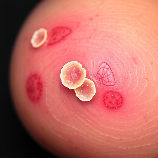
Under the magnifying lens of a dermatoscope, a common skin cancer can reveal far more than whether it is cancer at all — it can reveal how dangerous it is about to become. That is the central message of one of the largest investigations ever conducted into the surface signs of basal cell carcinoma, the most frequent malignancy in humans. Researchers at Ankara University in Turkey analyzed 1,091 histopathologically confirmed tumors removed from 690 patients between 2016 and 2023, and identified a short list of visible features that independently predict aggressive tumor biology hiding beneath the skin. Most strikingly, they found that ulceration — particularly when it involves more than half of the lesion’s surface — carries an odds ratio of 9.436, nearly a tenfold increase in the odds that a tumor harbors a high-risk subtype. The study, published in the Archives of Dermatological Research, suggests that the handheld lens many dermatologists already use in every examination could become a genuine forecasting tool rather than merely a diagnostic one.
Basal cell carcinoma arises from the basal cells of the deepest layer of the epidermis and accounts for the overwhelming majority of skin cancers diagnosed worldwide, with incidence climbing steadily as populations age and cumulative ultraviolet exposure mounts. The great majority of these tumors grow slowly, almost never metastasize, and are cured by straightforward excision. Yet the disease is far from uniform. Pathologists divide it into subtypes with very different temperaments: superficial and nodular variants generally remain confined and behave indolently, while micronodular, infiltrative, and basosquamous variants push thin strands and small nests of tumor cells deep into the dermis, can infiltrate along nerves, and recur more often after apparently complete removal. That distinction drives treatment intensity, because clinical guidelines match therapy to subtype: a superficial tumor may be managed with topical treatments or curettage, whereas an infiltrative tumor on the nose or ear may demand Mohs micrographic surgery with microscopic margin control. The problem is that these decisions are usually made on the evidence of a small punch or shave biopsy, and sampling error is a documented hazard. Previous studies cited by the Ankara team have shown that step sectioning of specimens initially diagnosed as superficial basal cell carcinoma frequently uncovers deeper, more aggressive components that the first thin slice missed.
Dermoscopy, also known as dermatoscopy or epiluminescence microscopy, bridges the gap between what the naked eye perceives and what the pathologist’s microscope later confirms. The technique employs a handheld device that magnifies the skin roughly tenfold and illuminates it with polarized light or through an immersion fluid, eliminating the white glare that normally bounces off the surface. Once surface reflection is suppressed, the examiner can look into the upper reaches of a lesion and observe structures invisible during ordinary inspection: the branching architecture of blood vessels, deposits of melanin at various depths, accumulations of keratin, and zones where the normal lattice of the skin has been erased. In basal cell carcinoma, the classic dermoscopic hallmarks — sharply focused arborizing vessels resembling bare tree branches, blue-gray ovoid nests and globules, leaf-like and spoke-wheel structures, and shiny white strands — allow trained examiners to distinguish these tumors from melanomas, benign moles, and inflammatory disorders with high accuracy. But as the Ankara authors point out, most earlier studies connecting dermoscopic patterns to histopathological subtype were limited by small sample sizes, particularly for the aggressive variants, leaving large-scale data on dermoscopic predictors of high-risk histology conspicuously scarce.
The new study was designed to close that gap. The team — dermatologists Dilan Kizilirmak and Bengu Nisa Akay, public health researcher Cemal Kocak, and pathologist Aylin Okcu Heper — assembled a retrospective cohort of every histopathologically confirmed basal cell carcinoma assessed at Ankara University’s Faculty of Medicine over an eight-year span. Each lesion was evaluated dermoscopically for vascular patterns, pigmentation, structureless areas, keratin, and ulceration using standardized terminology. The tumors were then sorted into a low-risk group, comprising superficial, nodular, and combined superficial-nodular lesions, and a high-risk group comprising micronodular, infiltrative, basosquamous, and mixed nodular-micronodular tumors. Multivariable logistic regression was then applied to determine which dermoscopic features remained independent predictors of high-risk histopathology once the influence of all other features was statistically controlled. The work was approved by the Ankara University Clinical Research Ethics Committee, conducted in accordance with the Declaration of Helsinki, and carried out with written informed consent from all patients.
The single strongest predictor was also the one most visible without any optical aid at all: ulceration. Ulcerated tumors — especially those in which the eroded surface covered more than half of the lesion — showed an odds ratio of 9.436 for high-risk histopathology, roughly a tenfold jump compared with lesions lacking the feature. The biological logic is compelling. Rapidly infiltrating carcinomas tend to outgrow their blood supply, and the resulting tissue death breaks through the epidermis as an ulcer; extensive ulceration therefore signals a tumor growing quickly and deeply enough to defeat its own nourishment. Ulceration has long been treated as a general warning sign in oncology, but the Ankara analysis quantifies, at unprecedented scale for this cancer, how strongly it flags aggressive biology in basal cell carcinoma — and shows that the signal intensifies with the area involved rather than depending on the mere presence of a small erosion.
Vascular architecture carried independent prognostic weight as well. Thick short serpentine vessels and long serpentine vessels — broader, more dilated, and more tortuous than the delicate vessels of indolent tumors — each independently predicted high-risk histopathology, while short fine serpentine vessels were markedly more frequent in low-risk lesions. Tumor angiogenesis offers a ready explanation: as a carcinoma invades deeper dermal layers, it recruits larger-caliber feeder vessels, and the caliber and course of vessels visible through the dermatoscope mirror the depth and vigor of that new blood supply. The behavior of blue-gray pigmentation told a parallel depth story. When blue-gray structures appeared sharply in focus — what dermoscopists call a focused pattern — the tumors were more often low-risk, consistent with small tumor nests sitting just beneath the epidermis, as in superficial basal cell carcinoma. When the blue-gray material looked blurred or unfocused, or combined focused and unfocused zones, high-risk histopathology predominated, reflecting tumor nests located deeper in the dermis, below the plane of crisp focus.
Keratin-related and white structural features added a further layer of discrimination. Isolated keratin — yellow-white, scale-like material in the absence of ulceration — and combinations of ulceration with keratin both independently predicted aggressive subtypes. Keratin production inside a basal cell tumor hints at squamous differentiation, the defining trait of basosquamous carcinoma, a hybrid malignancy sharing features of basal cell and squamous cell carcinoma that counts among the high-risk variants in the study’s classification. White calcified clods — bright, hard-edged white structures corresponding to dystrophic calcification within the tumor — and white circles, small round white structures that can mark keratin-filled invaginations, also emerged as independent predictors, as did small black clods. Each of these signs is uncommon enough that, viewed alone, it might be dismissed as a curiosity; the regression analysis demonstrates that, adjusted for the other features, they carry genuine statistical weight in anticipating what the pathologist will find.
Equally consequential is what the study says about reassuring signs. Pigmented structures and short fine serpentine vessels were significantly more frequent in low-risk lesions, meaning a tumor displaying these features and lacking the high-risk signals is more likely to belong to the indolent superficial or nodular families. The findings slot into a broader dermoscopic vocabulary for basal cell carcinoma assembled over two decades, ranging from the in-focus arborizing vessels of nodular tumors to the multiple aggregated yellow globules — known as MAY globules — recently linked to nonpigmented lesions. What the Ankara cohort adds is scale and confidence: with more than a thousand lesions, the statistical footing beneath each association far exceeds that of earlier series, which often rested on a few dozen aggressive cases. In practical terms, the study elevates several dermoscopic findings from anecdotal observation to evidence-based risk markers.
The clinical implications reach every step of the skin cancer pathway. When a dermatologist recognizes high-risk dermoscopic features before a biopsy is taken, the biopsy can be directed toward the most suspicious territory — the ulcerated rim, the zone of thick serpentine vessels — reducing the sampling error that can disguise an infiltrative tumor as a superficial one. Dermoscopy-guided site selection could make punch biopsies far more representative than they are today. Recognition of high-risk patterns before definitive treatment likewise informs management: tumors flagged dermoscopically as aggressive on the face or ears may be routed toward Mohs micrographic surgery or excision with careful margin assessment, while lesions carrying a low-risk signature may safely qualify for topical therapy, curettage, or simple excision, sparing patients more burdensome procedures. In telemedicine settings, where histopathology may be delayed, such a noninvasive risk readout could help prioritize which patients require urgent in-person evaluation, and the well-defined image features lend themselves naturally to future artificial-intelligence triage tools.
The authors are careful about the limits of their evidence. The study is retrospective and drawn from a single academic center, dermoscopic interpretation depends on examiner training, and a predictive model of this kind will need prospective validation in diverse populations before it can be woven into international guidelines. Even so, the central conclusion stands firm: dermoscopy delivers information that goes beyond the binary question of whether a lesion is cancerous and speaks to how it is likely to behave. As the researchers conclude, dermoscopy provides valuable risk stratification information beyond diagnosis alone, and specific vascular, keratin-related, and white structural features may help identify high-risk basal cell carcinoma while guiding both biopsy site selection and treatment planning. For a cancer expected to affect a large share of fair-skinned people during their lifetimes, a brief examination with a lens already hanging on the clinic wall now promises a preview not just of the diagnosis, but of the prognosis.
Subject of Research: Risk stratification of basal cell carcinoma using dermoscopic features to predict high-risk histopathological subtypes across 1,091 lesions.
Subject of Research: Medicine
Article Title: Risk stratification of basal cell carcinoma by dermoscopy: dermoscopic predictors from a large retrospective study of 1,091 lesions
Article References: Kizilirmak, D., Kocak, C., Heper, A. O., & Akay, B. N. (2026). Risk stratification of basal cell carcinoma by dermoscopy: dermoscopic predictors from a large retrospective study of 1,091 lesions. Archives of Dermatological Research, 318(1), Article 382. https://doi.org/10.1007/s00403-026-04854-7
Image Credits: AI Generated
DOI: 10.1007/s00403-026-04854-7
Keywords: basal cell carcinoma, dermoscopy, dermatoscopy, risk stratification, high-risk basal cell carcinoma, low-risk basal cell carcinoma, serpentine vessels, ulceration, MAY globules, histopathology, skin cancer
Cite Scienmag News
APA MLA Chicago
Rowan B. (August 29, 2026). Dermoscopy predicts basal cell carcinoma risk in study of 1,091 lesions. Scienmag. https://scienmag.com/dermoscopy-predicts-basal-cell-carcinoma-risk-in-study-of-1091-lesions/
Rowan B. “Dermoscopy predicts basal cell carcinoma risk in study of 1,091 lesions.” Scienmag, 29 August 2026, https://scienmag.com/dermoscopy-predicts-basal-cell-carcinoma-risk-in-study-of-1091-lesions/. Accessed 29 August 2026.
Rowan B. “Dermoscopy predicts basal cell carcinoma risk in study of 1,091 lesions.” Scienmag. August 29, 2026. https://scienmag.com/dermoscopy-predicts-basal-cell-carcinoma-risk-in-study-of-1091-lesions/
Copy citation Download RIS
Tags: Basal cell carcinoma diagnosisdermatological imaging for skin cancerDermatological research on tumor aggressivenessDermatoscopic features indicating high-risk BCCDermoscopy in basal cell carcinoma risk assessmentDermoscopy in skin cancerepidemiology of basal cell carcinomahandheld dermatoscope as a forecasting toolhigh-risk subtypes of basal cell carcinomaHistopathological confirmation of BCChistopathological confirmation of skin lesionslarge-scale skin cancer studyPredicting aggressive skin tumorsPrognostic indicators in basal cell carcinomaSkin cancer epidemiology and rising incidenceskin cancer prognosis using dermoscopySkin cancer risk assessment using dermoscopysurface features indicating tumor aggressivenessSurface features of basal cell carcinomasurface signs of aggressive skin tumorstumor surface ulceration and riskulceration as a predictor of high-risk BCCUlceration as a risk factor in BCCUse of handheld dermatoscope for skin cancer prediction