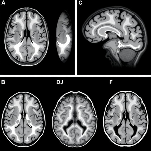
A new analysis of brain scans from hundreds of people has found that mild cognitive impairment (MCI), often considered an early stage on the path toward Alzheimer’s disease, can hide several biologically distinct patterns of brain change. The study, published in BMC Neuroscience, separates the overall amount of brain atrophy from the way damage is distributed across specific regions. That distinction revealed two structural subtypes of MCI with different relationships to memory, thinking and everyday functioning. The findings suggest that two people with similar levels of global brain shrinkage may nevertheless have substantially different anatomical profiles—and may experience different degrees of clinical impairment. By treating brain degeneration as both a widespread burden and a pattern of local changes, the researchers say, imaging studies may gain a more informative view of the complex transition between healthy ageing, MCI and Alzheimer’s disease.
MCI describes measurable problems with memory or other cognitive abilities that are greater than expected for a person’s age, but not always severe enough to prevent independent living. Some people with MCI remain stable for years, while others progress to Alzheimer’s disease or another dementia. This variability has made MCI difficult to study as a single biological category. Brain-imaging research has often attempted to divide patients into subgroups according to the regions that appear shrunken or otherwise altered. But those approaches can mix together two different signals: the overall severity of neurodegeneration and the local arrangement of structural abnormalities. A person with widespread, advanced atrophy may appear to have the same regional pattern as someone with milder global damage, even if the biological meaning of their scans is different. The new work was designed to disentangle those signals rather than allow one to obscure the other.
The researchers analysed baseline data from the Alzheimer’s Disease Neuroimaging Initiative, or ADNI, a large, multi-site programme that combines clinical assessments, brain imaging and other biological measurements. The dataset included 731 participants: 205 cognitively normal individuals, 351 people with MCI and 175 people diagnosed with Alzheimer’s disease. Structural magnetic resonance imaging was used to quantify brain anatomy. Unlike functional MRI, which tracks changes in blood flow or activity, structural MRI provides measurements of tissue volumes and regional morphology. The investigators first examined candidate brain regions across the full spectrum from healthy cognition to MCI and Alzheimer’s disease. This allowed them to identify anatomical measures that changed across the disease continuum before focusing specifically on the MCI group. The analysis used baseline observations, meaning that it was intended to characterise differences between participants rather than prove how an individual’s condition would evolve over time.
To estimate a participant’s overall neurodegenerative burden, the team created a global atrophy index, or GAI. The index was based on the ratio of total brain cerebrospinal-fluid volume to the combined volume of grey matter and white matter, with the tissue and fluid measures adjusted for total intracranial volume. The resulting values were then standardised as z-scores, which express how far an individual’s measurement lies from the average of the study population. In broad terms, more cerebrospinal fluid and less brain tissue indicate greater atrophy, because loss of neural tissue leaves additional space within the skull for fluid. Adjusting for total intracranial volume is important because skull size differs naturally among individuals and can otherwise distort comparisons. The researchers found that GAI followed a clear gradient: cognitively normal participants generally had the lowest global atrophy burden, people with MCI occupied an intermediate position, and participants with Alzheimer’s disease showed the greatest burden.
The crucial step came next. Within the MCI group, the investigators statistically removed the contribution of global atrophy from regional brain measurements, while also accounting for age, sex and education. This process, known as residualization, asks a specific question: after differences in overall atrophy and major demographic factors have been taken into account, which regional measurements remain unusually high or low? The residual is not a new physical measurement but the portion of a regional value that cannot be explained by the variables included in the model. This approach can expose local anatomical organisation that would otherwise be dominated by the general severity of brain loss. It also changes the interpretation of a small hippocampal, thalamic or frontal measurement: instead of simply marking that a brain is more atrophied overall, it may indicate that a particular network is disproportionately affected—or relatively preserved—compared with the person’s global condition.
The residualised measurements were then reduced into broader dimensions using principal component analysis, a mathematical technique that identifies combinations of variables explaining the greatest amount of variation in a dataset. Rather than considering every brain region independently, PCA can reveal coordinated patterns in which several areas change together. The researchers subsequently applied unsupervised clustering, which groups participants according to similarities in their imaging profiles without assigning them to categories in advance. Two principal local patterns emerged among people with MCI. One was characterised by lower residual measures involving the thalamus and striatum, structures deeply involved in information routing, movement, motivation and aspects of cognition. The other showed lower residual measures in frontal and paralimbic regions, including areas associated with executive control, emotional processing, memory and the integration of internal and external information. The labels describe anatomical patterns, not definitive disease mechanisms or diagnoses.
The two imaging-defined groups also differed clinically. Compared with the thalamic–striatal pattern, participants in the frontal–paralimbic subtype had greater cognitive impairment after statistical adjustment for relevant covariates. They also had poorer performance in measures of daily functioning. Cognitive status was assessed using the Alzheimer’s Disease Assessment Scale–Cognitive Subscale, commonly abbreviated ADAS-Cog13, a composite measure designed to capture problems in memory, language and other thinking skills. Everyday function was evaluated with the Functional Activities Questionnaire, or FAQ, which assesses difficulties with tasks such as handling finances, shopping, preparing meals and managing household responsibilities. The frontal–paralimbic subtype showed higher scores on both measures, with higher values indicating worse performance or greater impairment in these contexts. Associations with neuropsychiatric symptoms, however, were weak, suggesting that the structural distinction was more clearly related to cognition and practical function than to behavioural or psychological symptoms.
The researchers also found that continuous imaging features retained information that was lost when participants were assigned only to discrete subtypes. In statistical models predicting ADAS-Cog13 and FAQ scores, the PCA-derived residual features improved explanatory power beyond the cluster labels alone. This is a significant technical point: a category such as “frontal–paralimbic subtype” compresses a range of brain profiles into one group, whereas continuous scores preserve how strongly each individual expresses the underlying pattern. Two people can belong to the same cluster but differ considerably in the magnitude of their regional deviations. A model that includes those graded differences may therefore describe clinical variation more accurately than a binary or categorical label. The result does not mean that the imaging features can independently predict an individual’s future, but it indicates that the architecture of local brain changes contains measurable information about current cognitive and functional status.
The study’s broader implication is that global atrophy and local structural organisation should not be treated as interchangeable indicators of MCI. A global index can provide a useful estimate of how much neurodegeneration has occurred, while residual regional patterns may indicate which neural systems are particularly vulnerable relative to that burden. This framework could help researchers compare participants more fairly, refine clinical-trial populations and investigate why people with apparently similar levels of atrophy follow different clinical trajectories. It may also offer a way to integrate structural MRI with molecular biomarkers, genetic risk, cognitive testing and longitudinal outcomes. Yet the findings are not a clinical test, and they do not establish that either subtype is caused by a single pathological process. The analysis used publicly available, de-identified ADNI data and baseline measurements, so independent cohorts and follow-up scans will be needed to determine whether the patterns reliably predict progression to Alzheimer’s disease or other outcomes. For now, the work provides a more nuanced map of MCI—one that distinguishes the size of the problem from its anatomical shape.
Subject of Research: Brain-structure heterogeneity and clinically relevant imaging subtypes in mild cognitive impairment
Subject of Research: Medicine
Article Title: Disentangling global atrophy burden from local structural patterns reveals clinically relevant heterogeneity in mild cognitive impairment
Article References: “Disentangling global atrophy burden from local structural patterns reveals clinically relevant heterogeneity in mild cognitive impairment,” BMC Neuroscience
Image Credits: AI Generated
DOI: 10.1186/s12868-026-01035-0
Keywords: mild cognitive impairment, structural MRI, Alzheimer’s disease, global atrophy index, brain subtypes, residualization, principal component analysis, thalamus, striatum, frontal-paralimbic regions
Tags: Alzheimer’s disease early detection through brain imagingbiological subtypes of MCIbrain atrophy distribution and clinical impairmentbrain imaging biomarkers for Alzheimer’s diseasebrain imaging for predicting MCI progressionbrain region-specific damage in MCIbrain structural subtypes in cognitive declineclinical implications of brain atrophy patternsclinical significance of brain atrophy distributiondifferentiating MCI subtypes with brain scansdistinct brain degeneration subtypesdistinct brain region damage in MCIearly biomarkers of Alzheimer’s diseaseheterogeneity in MCI progressionheterogeneity of brain degeneration in MCIimpact of brain atimplications of brain imaging in neurodegenerative diseasesmild cognitive impairment brain atrophy patternsneuroanatomical profiles in mild cognitive impairmentneuroimaging of memory and thinking deficitsregional brain atrophy analysis in MCIstructural brain changes in cognitive declinetransition from healthy aging to dementia