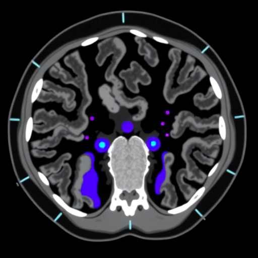
Computed tomography scans could become easier to interpret through a single artificial-intelligence-generated image that combines the information normally spread across several display settings, according to a study published in BMC Medical Imaging. The researchers developed a pipeline that converts routine Digital Imaging and Communications in Medicine, or DICOM, data into an “all-in-one” CT image designed to show soft tissue, lungs and bones simultaneously. In preliminary testing, the system produced images that closely matched its reference targets and enabled experts to detect most lung and bone lesions in a small clinical assessment. The approach could reduce the need for radiologists to switch repeatedly between different CT window settings, a routine step that adds time and cognitive burden to image interpretation.
A conventional CT scan contains far more information than is visible in any single screen display. CT attenuation is measured in Hounsfield units, a scale that distinguishes materials according to how strongly they weaken X-rays. Air-filled lung tissue occupies a very low range, soft tissues cluster around intermediate values, and dense bone lies at much higher values. Radiologists therefore adjust the window width and level—the range and midpoint of Hounsfield units mapped to grayscale—to emphasize particular anatomical structures. A lung window reveals faint differences in aerated tissue but can make soft-tissue contrast difficult to assess. A bone window highlights fractures and mineralized structures while suppressing many soft-tissue details. A soft-tissue window improves visualization of organs and masses but may obscure subtle lung or bone abnormalities. The new system attempts to preserve these complementary signals in one synthesized representation.
Creating a reliable target image for such a system is not straightforward because an all-in-one image is not directly acquired by the scanner. It is a secondary visual representation assembled from multiple window settings, and radiologists may disagree about how regions should be blended or prioritized. Instead of claiming to possess a definitive “ground truth,” the researchers constructed what they call a silver-standard reference. Their hybrid pipeline combined conventional image-processing operations with deep-learning segmentation to identify bone, lung and soft-tissue regions. Information from the different windows was then fused according to the segmented anatomy. The resulting images served as practical training references: not perfect physical measurements, but carefully engineered targets that encode the tissue-specific appearance clinicians need to examine.
The segmentation stage used TransU-Net, a model that combines the U-Net architecture’s ability to preserve fine spatial detail with transformer-based mechanisms capable of modeling relationships across a larger image. U-Net-like networks process an image through an encoder that extracts increasingly abstract features and a decoder that restores resolution, using skip connections to retain boundaries and local structure. Transformers add attention operations, allowing the network to weigh relationships between distant regions rather than relying only on neighboring pixels. That combination is useful in CT, where the identity of a structure may depend on its broader anatomical context. Tested on the public CT-ORG dataset, TransU-Net achieved a mean Dice similarity coefficient of 0.934. The Dice score measures overlap between predicted and reference regions, with 1 representing perfect agreement, indicating that the model could separate the principal anatomical categories with high consistency in the evaluation.
Once the regions had been identified and the silver-standard images generated, the investigators trained a Pix2Pix conditional generative adversarial network to perform the conversion directly from routine CT input. A GAN contains two competing neural networks: a generator creates an output image, while a discriminator attempts to distinguish generated images from reference images. During training, the generator learns not only to reproduce the overall intensity pattern but also to create images that appear structurally plausible to the discriminator. Pix2Pix is conditional because its output is guided by a corresponding input image rather than generated from random noise alone. In this study, that arrangement allowed the model to learn a mapping from standard DICOM-derived CT images to the fused all-in-one format. The researchers compared Pix2Pix with CycleGAN and AttentionGAN, two other image-translation approaches.
Pix2Pix produced the strongest numerical results among the tested models. Its structural similarity index measure, or SSIM, reached 0.99, suggesting close agreement in luminance, contrast and local structure with the reference images. Its peak signal-to-noise ratio was 43.42 ± 1.39 decibels, a high value indicating relatively low pixel-level reconstruction error. The model also recorded a Fréchet inception distance of 11.03 ± 8.64. FID estimates how closely the distribution of generated images resembles that of reference images in a feature space; lower values generally indicate greater similarity. These metrics do not prove that every clinically important abnormality has been preserved, but together they suggest that the generated images were visually and structurally close to the engineered targets. The results also illustrate why the reference-construction step matters: the model can only learn the tissue priorities and appearance conventions represented in the data used for training.
Expert reviewers found the synthesized images to have good overall quality and judged the depiction of abdominal, lung and bone structures favorably. The researchers then conducted a preliminary lesion-detection assessment using 64 follow-up CT examinations from patients with breast cancer. Accuracy was 85.94 percent for lung lesions and 87.50 percent for bone lesions. Those figures indicate that the generated images retained substantial diagnostic information in two clinically important categories, particularly because lung and skeletal abnormalities are often best appreciated in different conventional windows. The study does not establish that the AI-generated display is equivalent to a full radiologist review of the original scan, nor does it show that the system improves patient outcomes. Rather, it provides an early feasibility test of whether one synthesized image can preserve enough information to support lesion recognition.
The central technical challenge is balancing information compression against diagnostic fidelity. A single display must combine intensity ranges that are ordinarily separated because the grayscale dynamic range of a monitor is limited. Segmentation gives the algorithm a way to apply different transformations in different anatomical regions: lung information can be emphasized inside the thorax, bone detail can be enhanced around the skeleton, and soft-tissue contrast can be maintained in organs and masses. Yet anatomical boundaries are not always clean, and pathology can cross or distort the expected regions. A tumor, hemorrhage, atelectasis or treatment-related change may not conform to the categories used by a fusion rule. Generative models also learn statistical patterns from their training data, creating a need for careful validation to ensure that image synthesis does not smooth, distort or invent clinically relevant features. For that reason, the original CT data and conventional windows remain essential references rather than being rendered obsolete by the synthesized view.
The work was conducted by Yunjung Hong and Jiyoung Woo of Soonchunhyang University’s Department of ICT Convergence and Sanghyeok Lim of Soonchunhyang University Hospital Bucheon in the Republic of Korea. The retrospective hospital data were anonymized, and the study received institutional review-board approval; the public CT-ORG dataset was also anonymized. Supported by Soonchunhyang University and Korean National Research Foundation grants, the project points toward a possible change in how CT information is presented rather than how it is acquired. If validated across larger and more diverse datasets, an all-in-one display could help radiologists survey multiple tissue types more efficiently before examining suspicious regions in the original window settings. The authors describe the framework as a potential aid to multi-window interpretation, not an autonomous diagnostic system. Its next test will be whether the apparent visual convenience and strong reconstruction scores translate into reliable performance across scanners, diseases, institutions and real-world clinical workflows.
Subject of Research: Artificial-intelligence generation of all-in-one CT images from routine DICOM data using multi-window segmentation and image-to-image synthesis
Subject of Research: Medicine
Article Title: All-in-one CT generation from DICOM using hybrid multi-window segmentation, TransU-Net, and Pix2Pix
Article References: All-in-one CT generation from DICOM using hybrid multi-window segmentation, TransU-Net, and Pix2Pix — https://doi.org/10.1186/s12880-026-02711-7 Original publication
Image Credits: AI Generated
DOI: 10.1186/s12880-026-02711-7
Keywords: CT image generation, DICOM, TransU-Net, Pix2Pix, multi-window segmentation, generative adversarial networks, medical imaging, lesion detection
Tags: AI-assisted radiology interpretationAI-based enhancement of DICOM dataAI-powered CT image synthesisautomated multi-structure CT visualizationclinical evaluation of AI-generated CT imagescomprehensive CT visualizationDICOM to all-in-one CT conversionDICOM to all-in-one medical imaginghybrid segmentation in computed tomographyhybrid segmentation in medical imaginglesion detection in combined CT imageslung and bone lesion detectionmedical image enhancement with artificial intelligencemulti-organ visualization in medical imagingmulti-structure CT imaging techniquesPix2Pix for medical image translationPix2Pix image translation for radiologyradiology workflow optimization with AIreducing radiologist cognitive loadreducing radiologist workflow burdensoft tissue and bone simultaneous imagingTransUNet for medical image segmentation