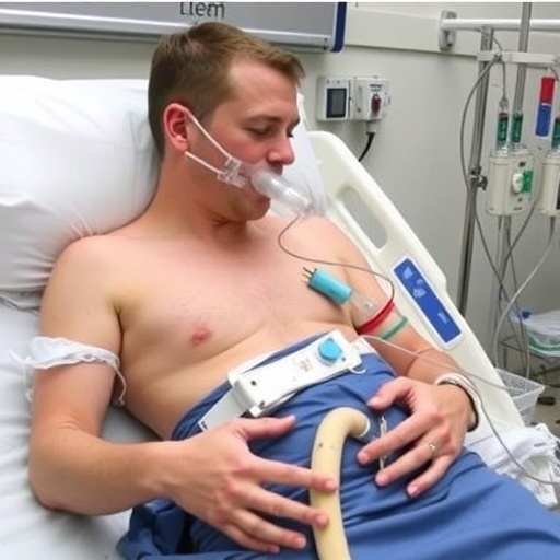
A new case report describes a “femoral-free” configuration for ambulatory venoarterial extracorporeal membrane oxygenation (V-A ECMO), a form of life support that can maintain circulation and oxygen delivery while allowing critically ill patients to participate in active rehabilitation. The technique, reported by surgeons at Keimyung University Dongsan Medical Center in South Korea, uses the right subclavian artery for arterial blood return and the right internal jugular vein for venous drainage. The approach is designed for patients who may require prolonged ECMO support, including those waiting for heart transplantation.
V-A ECMO is commonly used when the heart cannot pump enough blood to sustain the body, either because of severe cardiogenic shock, cardiac arrest, advanced heart failure, or failure after cardiac surgery. A typical peripheral setup places a large drainage cannula in a femoral vein and a return cannula in a femoral artery. Blood is removed from the venous system, passed through an oxygenator and pump, and then returned under pressure to the arterial circulation. Although effective, femoral cannulation often leaves patients confined to bed. The cannulas pass through the groin and upper leg, making standing, walking, and intensive physical therapy difficult and increasing the risk of complications associated with immobility.
Mobility is not a cosmetic benefit for patients on long-term ECMO. Prolonged bed rest can rapidly cause muscle wasting, weakness, loss of cardiovascular conditioning, pressure injuries, blood clots, and declining independence. For people being supported as a bridge to transplantation, preserving muscle strength and the ability to participate in rehabilitation may improve their chances of remaining eligible for surgery. The challenge is to provide reliable circulatory support without placing cannulas in locations that mechanically restrict movement or create unacceptable vascular risks.
In the reported case, the medical team initially attempted to provide arterial return through a vascular graft connected to the subclavian artery. The subclavian artery travels beneath the collarbone and supplies blood to the arm, while also offering a more upper-body access point than the femoral artery. However, the initial graft-based arrangement caused swelling in the patient’s upper limb. The problem highlighted the importance of preserving adequate blood flow through the arm and carefully managing the space occupied by the return circuit around the shoulder and chest.
The clinicians then modified the system. Instead of routing blood through a graft, they inserted a 17-French reinfusion cannula directly into the right subclavian artery. A 6-French distal perfusion catheter was added to maintain blood flow beyond the cannulation site and reduce the risk of arm ischemia. In practical terms, the distal perfusion catheter acts as a smaller auxiliary pathway, delivering blood to the downstream portion of the limb that could otherwise receive less circulation because of the larger arterial cannula. Monitoring the arm’s color, temperature, pulses, pressure, and tissue perfusion remains essential with this type of arrangement.
Venous drainage was established through the right internal jugular vein. The team used a long venous cannula and advanced its tip into the inferior vena cava, the large vessel that returns blood from the lower body to the heart. The cannula was introduced in a retrograde fashion, meaning it was advanced in the opposite direction from the conventional route often used for internal jugular venous access. Despite this unconventional orientation, the cannula reached a position that provided sufficient venous drainage for the ECMO circuit. Correct positioning is technically important because inadequate drainage can cause low circuit flow, excessive negative pressure, vessel collapse, or blood trauma.
With the revised configuration, the circuit maintained flows of approximately 3.5 to 4.0 liters per minute. Those flows were sufficient to provide stable V-A ECMO support in the reported patient while avoiding femoral cannulation. Most significantly, the patient was able to undergo active bedside rehabilitation. The arrangement left the legs and groin free, allowing movement that the authors say was comparable to the mobility possible with some dual-lumen veno-venous ECMO systems. Patients receiving dual-lumen veno-venous ECMO are often supported through a single large cannula in the neck, which can make mobilization more practical when the heart is still capable of providing adequate circulation.
The direct subclavian cannulation technique also appeared to resolve the arm-swelling problem observed with the original graft approach. No right-arm swelling was reported after the modification. Nevertheless, the authors emphasize that this finding comes from a single case and cannot establish that direct cannulation is safer or more effective than graft-based subclavian access. The subclavian artery is close to important nerves and structures in the chest and shoulder, and complications could include bleeding, thrombosis, vessel injury, infection, impaired limb perfusion, or difficulty controlling the cannulation site. The use of a distal perfusion catheter may reduce ischemic risk, but it does not eliminate the need for close surveillance.
The report is particularly relevant to bridge-to-transplant care, in which ECMO may be required for weeks or longer while a suitable donor organ becomes available. Conventional femoral V-A ECMO can be lifesaving, but the physical restrictions it imposes may contribute to a downward spiral of weakness and reduced transplant readiness. A configuration that permits earlier and more intensive physical therapy could help selected patients maintain strength while awaiting definitive treatment. It may also offer an alternative for individuals in whom femoral access is unavailable, unsuitable, or associated with excessive vascular risk.
However, the new approach should be viewed as a technical innovation rather than a validated replacement for standard peripheral V-A ECMO. The report provides experience from one patient, and the abstract does not establish long-term outcomes, comparative survival, rates of limb complications, or the durability of the cannulation sites. Larger case series and prospective studies would be needed to determine which patients are best suited for the technique, how it compares with axillary or femoral strategies, and whether the added complexity of direct arterial access translates into meaningful improvements in rehabilitation and transplant outcomes. For now, the case demonstrates that carefully planned upper-body cannulation can expand the possibilities for mobilizing patients who require prolonged mechanical circulatory support.
Subject of Research: Ambulatory venoarterial extracorporeal membrane oxygenation using right subclavian artery and right internal jugular vein cannulation as a bridge-to-transplant strategy.
Article Title: Ambulatory venoarterial ECMO via right subclavian artery and internal jugular vein cannulation: a novel technical approach to bridge-to-transplant
Article References: Jang, W. S., & Song, K. “Ambulatory venoarterial ECMO via right subclavian artery and internal jugular vein cannulation: a novel technical approach to bridge-to-transplant.” Journal of Artificial Organs, 29, Article 31 (2026).
Image Credits: AI Generated
DOI: 10.1007/s10047-026-01555-w
Keywords: Extracorporeal membrane oxygenation; ambulatory care; subclavian artery; internal jugular vein; heart transplantation; rehabilitation; bridge-to-transplant; artificial lung.
Tags: active rehabilitation during ECMO supportambulatory venoarterial ECMObridge-to-heart-transplant with ECMOcomplications of femoral cannulation in ECMOextracorporeal membrane oxygenation in cardiogenic shockfemoral-free ECMO configurationinnovative ECMminimally invasive ECMO techniquesmobility-friendly ECMO for critically ill patientsprolonged ECMO support for heart failuresubclavian and jugular cannulation for ECMOsustainable venoarterial ECMO setup