For decades, scientists have searched for ways to measure the tiny forces generated by living cells without disturbing the very behaviors they want to observe. Those forces can range from a few piconewtons—the scale of individual molecular interactions—to micronewtons produced by contracting cell groups. They shape how cells move, divide, attach to surfaces and respond to disease. Now, researchers have introduced an ultrasensitive “cell stethoscope” that uses an optical microcantilever to listen to the mechanical activity of living cells in real time. The device is designed to detect forces below one piconewton while operating in liquid, creating a new route for studying cellular mechanics under conditions closer to biology.
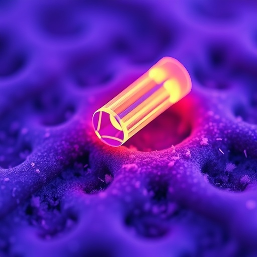
The technology, reported by Tu, Hao, Tu and colleagues in Nature Photonics, combines a miniature mechanical beam with an optical phase-sensing system built around a microfibre interferometer. A microcantilever is a small flexible structure that bends or vibrates when a force acts on it. In conventional force-sensing systems, the motion of such a beam may be monitored through changes in light intensity, electrical signals or laser position. The new approach instead tracks changes in the phase of light travelling through an interferometric structure. Because optical phase can respond to extremely small changes in distance or refractive conditions, it provides a highly sensitive way to translate mechanical motion into a measurable signal.
At the heart of the device is a microfibre-based interferometric structure integrated into the optical microcantilever. In an interferometer, light is divided into paths that later recombine. The resulting signal depends on whether the light waves arrive in step or out of step, a property known as phase. Even a minute displacement of the cantilever can alter the optical path length and shift this phase relationship. By monitoring that shift, the researchers can infer how the cantilever moves and calculate the force responsible. This method avoids relying on bulky external feedback arrangements and allows mechanical signals to be converted directly into high-resolution optical measurements.
The reported sensor achieves subpiconewton sensitivity and responses at megahertz-level frequencies, giving it both precision and speed. The combination is important because cellular forces are not always slow or steady. A cell may produce rapid changes as it contracts, attaches to a surface, responds to a chemical signal or interacts with neighboring cells. A sensor that is highly sensitive but too slow could miss these dynamics, while a fast sensor with insufficient resolution could fail to distinguish subtle mechanical events. The optical system described by the researchers is intended to capture both low-amplitude forces and fast fluctuations, making it suitable for observing mechanical behavior as it unfolds.
Operating in liquid is another central feature of the design. Many cellular experiments take place in aqueous culture media, but liquid environments create serious challenges for microcantilever sensors. Viscous drag can dampen motion, alter resonance behavior and reduce the quality of mechanical measurements. Conventional electronic or electromagnetic detection systems may also be vulnerable to interference from surrounding equipment. To address these problems, the researchers encapsulated the microfibre in a biocompatible film. The coating protects the optical structure from direct exposure to the liquid while preserving the sensor’s ability to respond to mechanical changes. It also supports compatibility with biological samples and helps shield measurements from electromagnetic interference.
The team demonstrated the device in liquid-phase physicochemical reactions, where it detected dynamic forces at piconewton resolution. Such tests are important because they show that the sensor is not limited to dry laboratory conditions or carefully isolated mechanical setups. Chemical reactions in liquids can generate changes in pressure, surface tension, molecular binding and fluid motion, all of which may produce small mechanical signals. Capturing those signals establishes a foundation for using the microcantilever in environments where biological processes occur. The results suggest that the platform can serve not simply as a force gauge, but as a general-purpose detector of mechanical activity in complex fluids.
The most biologically striking demonstration involved cardiomyocytes, the contractile muscle cells that generate the heartbeat. Under electrical stimulation, these cells rhythmically shorten and relax, producing mechanical forces that can be measured at their interface with the sensor. The cell stethoscope recorded the contractions quantitatively and in real time, while also monitoring their acoustic or vibrational signatures. In this context, “acoustic monitoring” refers to the detection of mechanical oscillations produced by cellular activity rather than ordinary audible sound. Measuring these signals could allow researchers to compare contraction strength, timing, rhythm and mechanical irregularities without destroying the cells or requiring them to be detached from their native environment.
The ability to study living cells non-invasively could be particularly valuable in research on cardiac disease, drug safety and regenerative medicine. Cardiomyocytes derived from stem cells are increasingly used to model inherited disorders and evaluate compounds that may affect heart rhythm. Electrical activity is often measured through voltage or calcium signals, but those readouts do not always reveal how strongly the cells contract. Mechanical force is a separate and essential dimension of cardiac function. By detecting contractions directly, the optical microcantilever could complement electrical and biochemical measurements, providing a more complete picture of how a treatment changes the performance of heart cells.
The transparent nature of the microcantilever adds another layer of capability. Because the structure permits optical observation of the cell beneath it, researchers can combine mechanical measurements with conventional microscopy and calcium fluorescence imaging. Calcium ions regulate contraction in cardiomyocytes, so fluorescent calcium indicators can reveal when intracellular calcium rises and falls. Simultaneously recording calcium activity and force production may expose mismatches between biochemical signaling and mechanical output. For example, a cell could exhibit normal calcium transients but weakened contraction, or produce force with abnormal timing. This multiparametric approach could help distinguish defects in signaling, force generation and mechanical coupling, while reducing the need for separate experiments.
The researchers present the cell stethoscope as a versatile platform for cellular mechanics across different environments, rather than as a tool restricted to cardiomyocytes. Similar measurements could ultimately be applied to cells that migrate, adhere, divide or remodel their surroundings. The sensor’s optical operation, liquid compatibility, electromagnetic immunity and high bandwidth address several long-standing limitations of microcantilever force probes. Challenges will remain before the technology becomes routine, including adapting the sensor to different cell types, calibrating measurements in complex biological fluids and integrating it into standardized laboratory workflows. Nevertheless, the combination of subpiconewton sensitivity, rapid response and simultaneous optical imaging offers a compelling new way to hear—and quantify—the hidden mechanical life of cells.
Subject of Research: A piconewton-sensitive optical microcantilever “cell stethoscope” for real-time, non-invasive measurement of cellular mechanical forces, including cardiomyocyte contractions.
Article Title: Piconewton-sensitive cell stethoscope using an optical microcantilever
Article References: Tu, X., Hao, G., Tu, W. et al. “Piconewton-sensitive cell stethoscope using an optical microcantilever.” Nature Photonics (2026). https://doi.org/10.1038/s41566-026-01964-6
Image Credits: AI Generated
DOI: https://doi.org/10.1038/s41566-026-01964-6
Keywords: Cell mechanics, optical microcantilever, piconewton force sensing, microfibre interferometry, cardiomyocyte contractions, cellular biomechanics, calcium fluorescence imaging, live-cell analysis, liquid-phase sensing
Tags: Cell mechanical force measurementinnovative cellular force monitoring devicesliquid environment cellular force measurementmicrofibre interferometer technologymicroscopic cell behavior analysisminimally invasive cellular force sensorsnanoscale force detection in living cellsoptical microcantilever biosensoroptical phase-sensing in biomechanicspiconewton-sensitive microcantileverreal-time cellular mechanicsultrasensitive biological force sensing