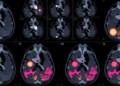
A new study in Nature Neuroscience reports that changes in the thickness of the cerebral cortex can appear at least seven years before amyloid reaches high levels in the brain, challenging the long-standing assumption that substantial amyloid accumulation is among the earliest detectable biological events in Alzheimer’s disease. The finding places subtle structural changes in the brain on an earlier point of the disease timeline and raises the possibility that neurodegenerative processes may begin well before amyloid becomes prominent enough to be identified by conventional thresholds. For a field increasingly focused on detecting Alzheimer’s decades before memory loss, the result could reshape how researchers define the earliest stages of the condition.
The cerebral cortex is the brain’s outer layer and contains densely organized networks responsible for memory, language, attention, perception, and decision-making. Its thickness reflects a combination of factors, including the size and arrangement of neurons, the complexity of their connections, supporting glial cells, and the organization of cortical layers. In healthy aging, cortical thinning occurs gradually, but Alzheimer’s disease is associated with more pronounced changes in specific regions, especially networks involved in episodic memory and higher-order cognition. Researchers measure these differences using high-resolution magnetic resonance imaging, which can estimate cortical thickness across thousands of points on the brain’s surface.
Amyloid-beta, meanwhile, is a protein fragment produced during the normal processing of amyloid precursor protein in neuronal membranes. Under pathological conditions, amyloid-beta can accumulate into plaques between brain cells. Positron emission tomography, or PET, allows researchers to visualize this accumulation using molecular tracers that bind to amyloid deposits. A person may show detectable amyloid years before symptoms develop, but the new report indicates that measurable cortical changes may precede the stage at which amyloid burden becomes high. This distinction is important because “amyloid-positive” is not a single biological state: the brain may pass through a prolonged period of low or intermediate accumulation before reaching levels associated with a more advanced disease process.
Roe, Jagust, Landau and colleagues examined the temporal relationship between cortical thickness and amyloid burden, focusing on whether structural alterations could be detected before amyloid crossed a high-level threshold. Their analysis, published under the title “Cortical thickness changes precede high levels of amyloid by at least 7 years,” suggests that the answer is yes. The wording of the study is deliberately precise. It does not claim that cortical thinning replaces amyloid as the defining biological marker of Alzheimer’s disease, nor does it establish that every early change in cortical thickness is caused by amyloid. Instead, it indicates that alterations in brain structure can become evident earlier than a later phase of substantial amyloid accumulation.
The timing is potentially transformative. A seven-year lead may provide researchers with a wider window in which to study the transition from biological risk to measurable brain injury. It could also influence the design of clinical trials, which increasingly seek volunteers before extensive neuronal damage has occurred. Treatments aimed at reducing amyloid may be most effective when administered before downstream degeneration becomes widespread. If cortical changes can identify individuals entering this vulnerable period, MRI-based measures could complement molecular biomarkers and help investigators determine when intervention should begin.
The result also adds complexity to the biological model of Alzheimer’s disease. The traditional amyloid cascade hypothesis proposes that amyloid accumulation initiates a sequence of events involving abnormal tau protein, inflammation, synaptic dysfunction, neuronal injury, and eventually cognitive decline. Modern versions of the model recognize that these processes overlap and influence one another rather than unfolding as a simple chain. Early cortical thinning could reflect several mechanisms, including synaptic loss, altered neuronal metabolism, vascular contributions, inflammation, or changes related to tau pathology. It might also capture individual differences in brain development, reserve, or vulnerability that are not visible in amyloid measurements alone.
That does not mean that MRI can currently diagnose Alzheimer’s disease seven years in advance. Cortical thickness varies naturally between individuals, and normal aging can produce regional thinning without leading to dementia. Measurements are also sensitive to scanner hardware, image quality, processing pipelines, statistical models, and the choice of brain regions examined. A robust clinical test would need to distinguish disease-related change from normal variation and from other conditions that affect the cortex, including cerebrovascular disease, frontotemporal degeneration, traumatic brain injury, and psychiatric or neurological disorders. The study’s significance therefore lies less in offering an immediate screening tool than in showing that brain structure may contain an earlier signal than previously appreciated.
The findings may become especially valuable when combined with other biomarkers. Amyloid PET can reveal plaque-related pathology, while tau PET provides information about the distribution of abnormal tau, and cerebrospinal fluid or blood tests can measure proteins associated with amyloid, tau, neuronal injury, and inflammation. MRI adds a different layer of information: it shows how the brain’s architecture is changing. Integrating these modalities could allow researchers to build more detailed disease trajectories, identifying whether cortical thinning appears before amyloid becomes high, alongside emerging tau pathology, or in association with other biological processes. Such models could improve risk prediction while helping scientists separate the earliest causes of degeneration from later consequences.
For the public, the message is both striking and cautionary. The brain may begin changing long before memory problems become noticeable, but an early structural signal is not the same as a certain prediction of dementia. The study instead reinforces the idea that Alzheimer’s disease is a prolonged biological process, not an event that begins when symptoms first appear or when a single scan turns positive. Its most important contribution may be conceptual: cortical structure deserves attention as an early indicator of brain vulnerability, while amyloid burden should be interpreted as one component of a much larger and evolving disease picture. As researchers continue mapping the sequence of changes that precede cognitive decline, the earliest years of Alzheimer’s biology are becoming visible—and potentially actionable—long before the disease announces itself in everyday life.
Subject of Research: Early brain changes associated with Alzheimer’s disease, including cortical thickness and amyloid accumulation.
Article Title: Cortical thickness changes precede high levels of amyloid by at least 7 years.
Article References: Roe, J.M., Jagust, W.J., Landau, S.M. et al. “Cortical thickness changes precede high levels of amyloid by at least 7 years.” Nature Neuroscience (2026). https://doi.org/10.1038/s41593-026-02363-4
Image Credits: AI Generated
DOI: https://doi.org/10.1038/s41593-026-02363-4
Keywords: Alzheimer’s disease, cortical thickness, amyloid, brain imaging, magnetic resonance imaging, PET, neurodegeneration, early detection, biomarkers, cognitive decline
Tags: advances in neuroimaging for Alzheimer’sAlzheimer’s disease early detectionamyloid and brain structure relationshipbrain structural changes before amyloid accumulationcortical thickness measurementcortical thinning in neurodegenerationearly Alzheimer’s diagnosisneuroimaging in Alzheimer’spreclinical biomarkers of Alzheimer’srole of cerebral cortex in cognitive declinesignificance of cortical thinning in dementiatimeline of Alzheimer’s disease progression