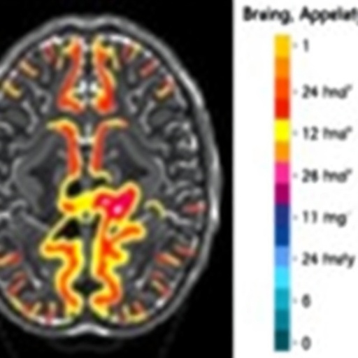
A new brain-imaging approach could give researchers a more sensitive way to track Huntington’s disease, revealing microscopic changes in brain tissue before they are fully captured by conventional measurements of brain volume. In a study published in eLife, scientists used a diffusion magnetic resonance imaging model known as Soma and Neurite Density Imaging, or SANDI, to detect abnormalities in the brains of people living with the inherited neurodegenerative disorder. The findings suggest that the technique may eventually help clinical trials determine whether experimental treatments are slowing or preventing damage to brain cells.
Huntington’s disease is caused by an inherited mutation and gradually damages regions of the brain involved in movement, learning, habits and cognition. The disease is especially destructive in the basal ganglia, a network of deep-brain structures that includes the striatum. Neurons in the striatum, particularly medium spiny neurons, are among the earliest cells to degenerate. As these cells and supporting tissue are lost, the striatum shrinks, contributing to worsening motor control and cognitive function. Although several disease-modifying therapies are being tested, researchers still need reliable, non-invasive biomarkers that can show how the disease is progressing inside the brain.
Standard MRI scans can measure the volume of brain structures and are already widely used in Huntington’s disease research. However, volume measurements provide a relatively broad view of tissue loss. They do not directly reveal the microscopic changes that cause atrophy, such as the disappearance of neurons, alterations in supporting glial cells or changes in the spaces between cells. SANDI was developed to provide a more detailed perspective. It analyses the movement of water molecules through tissue during a diffusion MRI scan, using mathematical modelling to estimate features such as the size and density of cell bodies, known as somas, as well as the density of neurites, the slender projections extending from neurons.
Researchers at Cardiff University Brain Research Imaging Centre tested the model using MRI data from 56 people with Huntington’s disease and 57 healthy volunteers. All participants were scanned on the same MRI system, helping to reduce technical differences that can complicate comparisons between hospitals and imaging centres. Participants with Huntington’s disease also completed motor assessments, including rapid and paced finger-tapping tasks. These tests allowed the investigators to examine whether microscopic imaging signals were related to measurable problems with movement.
The team compared SANDI-derived measurements in the striatum with measurements from the thalamus, another deep-brain structure. The thalamus was selected as a control region because neurodegeneration in Huntington’s disease typically begins in the striatum and spreads to neighbouring areas, including the thalamus, later in the disease course. This design enabled the researchers to ask whether SANDI could identify changes in a region expected to be affected early while showing fewer abnormalities in a region expected to be relatively preserved at that stage.
The results revealed a distinctive pattern in the striatum of people with Huntington’s disease. Estimated soma density was lower than in healthy volunteers, while estimated soma size and the amount of space between cells were higher. The researchers say this combination is consistent with the loss of neurons and the accompanying reaction of glial cells, which support and protect neurons but can change in number and structure during neurodegeneration. The imaging pattern therefore appeared to correspond with microscopic abnormalities previously documented in post-mortem studies of Huntington’s disease.
When combined with participants’ age, the SANDI measurements explained as much as 63 percent of the shrinkage observed in the striatum. The same imaging indices were also associated with poorer performance on the finger-tapping tasks, linking the estimated tissue abnormalities to functional motor impairment. By contrast, the researchers did not observe comparable changes in the thalamus. This regional distinction strengthens the possibility that SANDI is detecting disease-related biology rather than simply reflecting general differences in brain size or image quality.
The findings do not yet establish that SANDI can track disease progression better than existing biomarkers. The study was based on a single imaging session, and larger, long-term investigations will be needed to determine whether the measurements change consistently as Huntington’s disease advances. Researchers must also test how well the model performs across different MRI scanners and standard hospital imaging systems. Such validation is essential before SANDI can be used routinely in clinical trials or patient care.
Even with those limitations, the technique could provide a valuable layer of information alongside conventional volumetric MRI. A treatment might preserve brain volume only modestly while still producing measurable effects on cellular structure, or microscopic improvements might appear before changes in overall anatomy become detectable. By estimating tissue properties associated with neuronal loss and glial responses, SANDI could help scientists assess whether an experimental therapy is protecting vulnerable brain cells. The approach may also be adaptable to other neurodegenerative conditions, including Alzheimer’s and Parkinson’s diseases, although each disorder will require separate validation. The study, led by Vasileios Ioakeimidis and senior author Claudia Metzler-Baddeley, presents SANDI as a promising tool for turning advanced MRI into a closer window on the cellular damage driving Huntington’s disease.
Subject of Research: People
Article Title: In vivo mapping of striatal neurodegeneration in Huntington’s disease with Soma and Neurite Density Imaging
News Publication Date: 11-Aug-2026
Web References: https://elifesciences.org/; https://doi.org/10.7554/eLife.107661.3
References: Ioakeimidis et al., “In vivo mapping of striatal neurodegeneration in Huntington’s disease with Soma and Neurite Density Imaging,” eLife, DOI: 10.7554/eLife.107661.3
Image Credits: Ioakeimidis et al. (CC BY 4.0)
Keywords: Huntington’s disease, neurodegeneration, brain imaging, magnetic resonance imaging, diffusion MRI, SANDI, soma density, striatum, basal ganglia, disease progression, neurological disorders, clinical trials
Tags: advanced MRI methods for brain structure analysisbasal ganglia and striatum in Huntington’s diseaseclinical trial assessment of neurodegenerative treatmentsdiffusion magnetic resonance imaging in neurodegenerationearly diagnosis of neurodegenerativeHuntington’s disease early detectionmicroscopic brain tissue changes in neurodegenerative disordersNeurodegenerative disease brain imagingnon-invasive biomarkers for neurodegenerative progressionSoma and Neurite Density Imaging (SANDI) techniquetracking brain tissue degeneration in Huntington’s disease