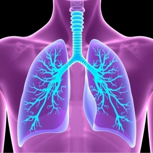
Lung cancer remains the world’s most commonly diagnosed cancer and one of its deadliest diseases, but a new artificial intelligence system could help physicians reach tumors that have long been difficult to access. Developed by researchers at Pusan National University in South Korea, ASTRA-Net is designed to reveal tiny peripheral airways that are frequently missing from computed tomography (CT) airway maps. By reconstructing these overlooked branches, the system may provide a more complete route for navigational bronchoscopy, a procedure used to guide instruments deep into the lungs for diagnosis and treatment.
Obtaining tissue from small lung lesions is essential for determining whether a suspicious growth is cancerous, yet the procedure can be technically demanding. Many nodules develop near the outer regions of the lung, far from the larger airways through which bronchoscopic instruments enter. To reach them, clinicians must navigate an intricate, tree-like network of increasingly narrow passages. Modern robotic and computer-assisted bronchoscopy systems rely on three-dimensional airway maps generated from CT scans, but these maps are only as useful as the airway structures they contain.
The smallest airways are particularly difficult to identify in CT images. Their diameters may approach the limits of imaging resolution, while surrounding tissues, blood vessels, motion artifacts, and variations in image quality can obscure their boundaries. Manual annotation is also time-consuming, meaning that some peripheral branches may be unintentionally left out of the labels used to train an AI model. If an algorithm learns exclusively from these incomplete annotations, it may reproduce the omissions rather than discover the anatomy that was missed.
ASTRA-Net, short for Anatomical Segmentation with Tree-aware Refinement Attention, was created to address this problem. Rather than treating the existing labels as a complete description of the lung airway tree, the framework is designed to identify anatomically plausible structures beyond the annotated regions. Its architecture combines broad anatomical segmentation with targeted refinement. One component first learns the overall organization of the lungs and their major airways, while another concentrates on areas where airway boundaries are poorly defined or where small branches may have been overlooked.
The model’s attention mechanism helps it prioritize regions that are especially challenging to interpret. In these areas, ASTRA-Net analyzes more than isolated image intensity patterns. It considers the continuity of airway pathways and uses anatomical relationships within the surrounding lung. Blood vessels, which often travel alongside airways, can provide additional contextual clues. By combining these signals, the system can infer whether a faint structure represents a continuation of an existing airway rather than random imaging noise or an unrelated anatomical feature.
This tree-aware strategy is important because the bronchial system is not a collection of independent tubes. It is a connected branching network in which each airway follows patterns of anatomical continuity. A model that recognizes those relationships may be better equipped to distinguish a plausible distal branch from a false prediction. ASTRA-Net therefore seeks not only to improve pixel-level segmentation, but also to preserve the topology of the airway tree—the way branches connect, divide, and extend toward the lung periphery.
Researchers evaluated the framework on multiple datasets, including clinical CT scans collected at Pusan National University Yangsan Hospital. The system showed strong performance in detecting fine peripheral airways and maintained its effectiveness across scans with different image qualities and slice thicknesses. These variations are clinically significant because CT protocols differ between hospitals and patients. An algorithm that performs well only on highly standardized images may have limited value in everyday practice, where scans can contain noise, thicker sections, or reduced contrast.
One of the study’s most revealing findings emerged during expert review. Some structures initially classified as false positives by conventional evaluation methods were judged by specialists to be genuine airway branches absent from the original annotations. This highlights a central difficulty in training and testing medical AI: a model can appear to make errors when the reference labels are incomplete. In ASTRA-Net’s case, predictions that disagreed with the annotations were not necessarily incorrect; some may have represented anatomy that the labels failed to capture.
The researchers say a more complete airway roadmap could help physicians plan routes to difficult-to-reach lesions and support future AI-assisted or robotic bronchoscopy systems. By extending airway maps farther into the lung, ASTRA-Net may improve the ability of navigation platforms to identify possible paths before a procedure begins, potentially reducing uncertainty during tissue sampling. The technology is not presented as a replacement for clinical judgment, and further validation will be needed to determine how reconstructed airways perform in real-time procedures and whether they improve diagnostic outcomes. Nevertheless, the work points toward a broader shift in medical imaging: AI systems may become capable not only of reproducing expert annotations, but also of detecting clinically meaningful structures that those annotations missed.
Subject of Research: Experimental study
Article Title: Discovery of Peripheral Airway Beyond Incomplete CT Annotations for Navigational Bronchoscopy
News Publication Date: 1-Jun-2026
Web References: https://doi.org/10.1109/TMI.2026.3672178
References: IEEE Transactions on Medical Imaging, DOI: 10.1109/TMI.2026.3672178
Image Credits: Professor MinWoo Kim, Pusan National University, Korea
Keywords: ASTRA-Net, artificial intelligence, medical imaging, lung cancer, airway segmentation, computed tomography, CT scans, navigational bronchoscopy, peripheral airways, robotic bronchoscopy, biomedical engineering, pulmonary medicine
Tags: advanced pulmonary diagnostic toolsAI-assisted lung navigationASTRA-Net artificial intelligence systemcomputerized tomography lung imagingCT airway map enhancementlung airway mappinglung cancer detection technologyminimally invasive lung biopsynavigational bronchoscopy guidanceperipheral airway reconstructionsmall airway visualizationsmall lung lesion diagnosis