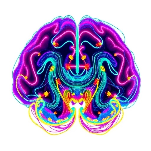
Nanoplastics—plastic fragments smaller than one micrometer—are now detected across food, water, air, and even biological tissues. Yet understanding how these particles travel inside complex organs, especially the brain, remains a major scientific hurdle. Traditional methods often require thin tissue sections, limiting observations to two dimensions and making whole-organ mapping difficult.
A new study by researchers at Japan’s National Institute for Environmental Studies (NIES), the University of Osaka, and Waseda University addresses this gap with a workflow designed for three-dimensional brain visualization without sectioning. The focus is the neonatal period, when the brain is rapidly developing and biological barrier systems are still immature.
The team used fluorescently labeled polystyrene nanoplastics and delivered them orally to neonatal mice. Twenty-four hours later, brains and other organs were collected and imaged. The results reveal a strong size-dependent biodistribution: 50 nm particles accumulated far more extensively than larger 500 nm particles, not only in the brain but also in the intestine and kidneys.
To visualize this distribution throughout an intact organ, the researchers applied a tissue-clearing protocol (SeeDB2G) to render neonatal brain tissue optically transparent. This step enabled fluorescence imaging deep inside the tissue while preserving spatial structure.
Light-sheet fluorescence microscopy then provided whole-brain three-dimensional reconstructions. Rather than reporting only regional “presence,” the approach allowed signal intensity comparisons across the brain’s internal architecture.
Quantitative analysis indicated relatively higher fluorescence in the thalamus and brainstem compared with regions such as the cerebral cortex and cerebellum. Because these areas lie close to the ventricular system, the pattern may relate to cerebrospinal fluid circulation or developmental changes in early barrier properties.
Importantly, the study validates its fluorescence readouts using hyperspectral imaging, helping distinguish particle-derived signal from potential confounders such as free dye leakage or tissue autofluorescence. The spectral signatures matched those expected for polystyrene nanoplastics, strengthening confidence in the observed localization.
While the work demonstrates a powerful mapping capability—and suggests where nanoplastics may preferentially accumulate—it does not assess environmentally realistic exposure levels or directly evaluate health risks. Its contribution is methodological: establishing a platform for future studies on entry routes, circulation dynamics, and clearance mechanisms.
Subject of Research: Animals
Article Title: Whole-Tissue Distribution Analysis for Visualization of Nanoplastics in the Neonatal Mouse Brain
News Publication Date: 29-Jun-2026
Web References: http://dx.doi.org/10.1016/j.hazadv.2026.101353
References: 10.1016/j.hazadv.2026.101353
Image Credits: National Institute for Environmental Studies (NIES)
Keywords: nanoplastics, biodistribution, neonatal brain, tissue clearing, light-sheet fluorescence microscopy, environmental toxicology, nanoparticles, central nervous system
Tags: 3D tissue clearing and imaging of nanoplasticsenvironmental nanoplastic contamination andfluorescently labeled nanoplastics in biological tissuesimpact of nanoplastics on neonatal healthlight-sheet fluorescence microscopy in neurosciencenanoplastic detection in biological tissuesNanoplastic distribution in neonatal mouse brainsnanoplastic penetration in developing organsneonatal brain development and nanoplastic exposuresize-dependent nanoplastic biodistributiontissue-clearing protocols for organ visualizationwhole-organ 3D mapping of nanoplastics