Blood vessels must form in a way that is both precise and reliable, so that oxygen and nutrients can reach every part of the body. During development, endothelial cells organize into a branched network of hollow tubes, creating a continuous lumen that later allows blood to flow. Yet how neighboring vessel segments coordinate their movement and connect into an unbroken channel has remained only partially understood.
Using high-resolution live imaging in zebrafish, researchers from the University of Basel—led by Professor Markus Affolter and Dr. Heinz-Georg Belting—mapped the process with unprecedented detail. Their work, published in eLife, reveals the full sequence by which adjacent lumina merge to generate mature, connected blood vessels.
The study focuses on the junctions that keep endothelial cells attached to one another during tube formation. These cell-cell contacts must be stable enough to preserve the integrity of the vessel wall, while also remaining adaptable to permit rearrangements of the cells.
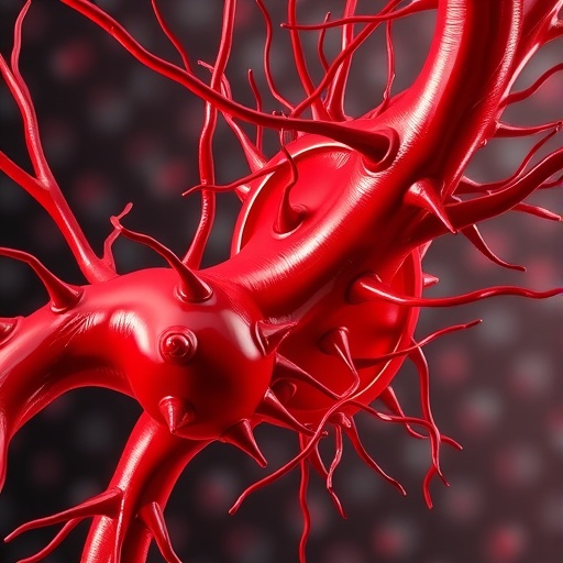
A key player in this remodeling is junction-based lamellipodia (JBL), specialized membrane protrusions that drive endothelial cell repositioning. The new findings explain the mechanical logic behind how these JBL structures enable cells to extend, meet, and link.
The process begins when a JBL protrusion forms at the tip of an endothelial cell, producing a pushing force that advances the cell front. At the protrusion’s tip, the junction acts like a molecular anchor, attaching to a neighboring cell and stabilizing the growing interface between segments.
Then pulling forces take over, drawing the rear of the cell forward and gradually elongating the cell. In repeated push-and-pull cycles, cells behave in an inchworm-like manner, progressively extending the lumen until separate segments fuse into a continuous vessel.
The researchers report that VE-cadherin, a major junction molecule, plays two distinct roles: it provides “glue” for adhesion and vessel-wall stability, and it actively supports cell movement. Notably, endothelial cells appear both robust and highly plastic during the same developmental steps.
Beyond clarifying vascular development, the work suggests general principles for how tissues could be patterned around working blood supply. Because many organs develop in close coordination with vasculature, improved understanding may translate into better strategies for engineering vascularized tissues.
In the long term, these insights could help create organoids and laboratory-grown tissues that include functional blood vessels—an essential step toward making regenerative medicine more faithful to natural development.
Subject of Research: Endothelial cell mechanics and junction-mediated vascular tube formation
Article Title: Junctional and Actomyosin Dynamics Drive Endothelial Cell Rearrangements during Vascular Tube Formation.
Web References: http://dx.doi.org/10.7554/eLife.109264.2
References: eLife, 10.7554/eLife.109264.2
Image Credits: Etienne Schmelzer, Biozentrum, University of Basel
Tags: blood vessel formationblood vessel morphogenesis mechanismscell-cell adhesion in blood vessel developmentendothelial cell junction remodelingendothelial cell migration and connectionforces driving blood vessel fusionhigh-resolution imaging of vascular tissuesjunction-based lamellipodia in blood vesselslive imaging of blood vessel developmentvascular network patterningvessel lumen formationzebrafish models in vascular research