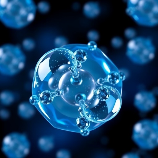
Lipid-shelled nanobubbles are emerging as a versatile upgrade to ultrasound contrast, aiming to overcome limits imposed by weak contrast resolution in many clinical scans. While today’s routine agents rely on micron-scale microbubbles—effective but comparatively large—nanobubbles promise greater flexibility for disease-specific detection and, potentially, combined diagnostic and therapeutic functions.
The new work details a practical, step-by-step laboratory procedure for formulating and activating lipid-shelled nanobubble ultrasound contrast agents. According to the study, the typical workflow uses self-assembly driven by mechanical agitation, followed by purification via differential centrifugation to enrich nanobubbles and reduce unwanted microbubble contamination.
A central technical theme is that the formulation is “deceptively simple” yet highly sensitive to minor deviations. Parameters such as mixing conditions, assembly timing, and centrifugation settings can influence the resulting size distribution and therefore the bubble’s acoustic behavior—directly affecting how strongly the agent responds to ultrasound.
The protocol also covers how different variants can be engineered from the same foundational platform: plain nanobubbles, fluorophore-conjugated versions for optical tracking, ligand-conjugated constructs for targeting pathological markers, hydrophilic dye-integrated formulations for imaging contrast, and drug-loaded bubbles designed for therapeutic delivery.
Because lipid composition governs membrane stability, careful handling is required during activation to preserve shell integrity. The authors emphasize that poor reproducibility—stemming from small procedural errors—can translate into measurable changes in yield, stability, and acoustic performance between batches.
For translational relevance, the study frames nanobubbles as particularly suited to diseases involving abnormal vasculature, where targeted accumulation could enhance sensitivity and specificity. The ability to decorate the bubble surface with functional moieties also supports multiplexed applications, from imaging biomarkers to delivering payloads.
In terms of feasibility, the procedure is designed for users with basic laboratory expertise and is reported to take about three hours to complete the preparation and activation steps for plain nanobubbles.
Overall, the protocol provides the technical guardrails needed to reliably produce lipid-shelled nanobubbles—supporting the broader development of next-generation ultrasound contrast beyond conventional microbubble formulations.
Subject of Research: Ultrasound contrast agents; lipid-shelled nanobubbles; formulation and activation protocols.
Article Title: Formulation and activation of lipid-shelled nanobubble ultrasound contrast agents.
Article References: Cooley, M.B., Kosmides, T., Nittayacharn, P. et al. Formulation and activation of lipid-shelled nanobubble ultrasound contrast agents. Nat Protoc (2026). https://doi.org/10.1038/s41596-026-01411-4
DOI: https://doi.org/10.1038/s41596-026-01411-4
Tags: centrifugation techniques for nanobubble purificationchallenges in reproducibility of nanobubble productioninfluence of formulation parameters on acoustic responselipid membrane stability in nanobubblesLipid-shelled nanobubblesnanobubble activation protocolsnanobubble customization with fluorophores and ligandsnanobubble formulationnanobubbles for therapeutic deliverytargeted nanobubbles for disease detectionultrasound contrast agentsultrasound imaging enhancement