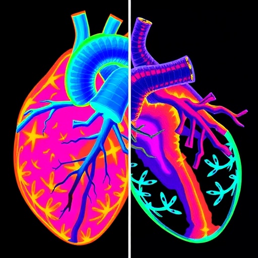
A new study is shining a microscope-sized flashlight into the heart’s inner architecture, using a technique that maps tissue mechanics at the voxel scale. The work, published in Nature Biomedical Engineering, tackles a long-standing problem in cardiovascular imaging: how to quantify microstructural remodeling in living human myocardium, not just in animal models or at coarse anatomical resolution, but throughout the whole heart.
Researchers led by Rock and colleagues applied diffusion tensor phenomapping to compare healthy hearts with those subjected to pressure overload. Pressure overload—such as that caused by chronic hypertension or valve disease—reshapes cardiac muscle over time, altering fiber organization, cellular alignment, and the way water molecules move through tissue.
Diffusion tensor imaging (DTI) exploits the fact that water diffusion inside myocardium is directionally constrained by the underlying fiber architecture. From DTI, scientists derive diffusion tensors and related metrics that can be interpreted as proxies for microstructural anisotropy and tissue organization. What makes the new approach stand out is the “phenomapping” layer: the team converts these diffusion characteristics into spatially resolved phenotypes that can be compared across the entire heart.
Using voxel-scale mapping, the authors reveal how diffusion signatures shift in pressure-overloaded tissue. The remodeling is not treated as a single global change; instead, it appears as region-specific alterations consistent with a breakdown and reorganization of normal myocardial structure. Such spatial heterogeneity matters because disease progression often begins locally, then propagates through mechanics and signaling.
The researchers’ comparisons between healthy and pressure-overloaded hearts support the idea that diffusion-derived descriptors can serve as quantitative biomarkers of remodeling. This could help link imaging patterns to underlying tissue-level processes such as altered fiber orientation and compromised microstructural integrity—details that are difficult to capture with conventional clinical imaging.
Importantly, the method remains grounded in physics-based diffusion measures while delivering a more interpretable phenotype map. For clinicians and bioengineers, that combination could accelerate how imaging translates into mechanistic hypotheses and, ultimately, risk stratification.
If validated in broader cohorts, voxel-scale diffusion phenomapping may become a viral, game-changing tool in the ongoing race to make heart disease visible before irreversible functional decline.
Subject of Research: Human heart remodeling under pressure overload using voxel-scale diffusion tensor phenomapping
Article Title: Voxel-scale diffusion tensor phenomapping of the healthy and pressure-overloaded human heart
Article References: Rock, C.A., Chen, Y.I., Wang, R. et al. Voxel-scale diffusion tensor phenomapping of the healthy and pressure-overloaded human heart. Nat. Biomed. Eng (2026). https://doi.org/10.1038/s41551-026-01755-y
Image Credits: AI Generated
DOI: https://doi.org/10.1038/s41551-026-01755-y
Keywords: (not provided)
Tags: cardiac fiber organization analysisdiffusion MRI in heart diseasediffusion tensor phenomapping in cardiovascular imagingin vivo heart tissue microarchitecturemicrostructural anisotropy in myocardiummyocardial microstructural remodelingpressure overload effects on heart tissuestructural changes in hypertensive heartstissue mechanics mapping in heart healthvoxel-scale diffusion tensor imagingwater diffusion constraints in cardiac tissuewhole-heart microstructure comparison