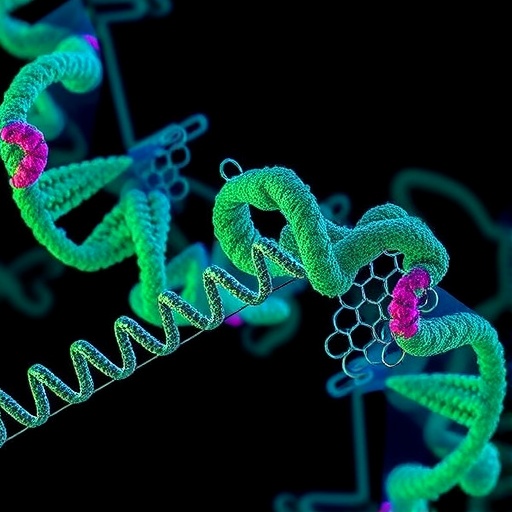
Trypanosome mitochondria rely on an RNA-editing process that rewrites transcripts through precisely timed uridine deletions and insertions, guided by complementary RNAs. For decades, researchers have known the editing is executed by a fast-moving, multi-enzyme system—but how the components coordinate their actions has been difficult to pin down.
Now, Liu and colleagues provide structural snapshots of two key editing machines by defining “editosomes” as supramolecular assemblies built from the RNA substrate-binding complex (RESC) together with either the RNA-editing catalytic complex 1 (RECC1) or complex 2 (RECC2). Using cryo–electron microscopy, they report approximately 1-MDa structures that reveal how the editing cascade is organized at near-atomic resolution.
The complexes adopt a striking architecture reminiscent of dragonflies. Each editosome includes a head-like region, a thorax-like core, and a tail and wing arrangement that correspond to distinct functional stages. RECC1 and RECC2 specialize in different chemical steps: the deletion cascade and the insertion cascade, respectively.
At the heart of each complex is a tetrameric core containing one active and three inactive RNase III domains. This core captures the guide RNA (gRNA)–mRNA duplex, creating a controlled substrate platform. Zinc-finger elements then discriminate between deletion and insertion sites, positioning the RNA for cleavage with site-specific accuracy.
Once the substrate is cleaved, peripheral modules reshape the reaction environment into a modular “reaction chamber.” Three oligonucleotide-binding-fold heterotetramers are flexibly attached to the core, suggesting that the machinery can adapt to different RNA geometries during the editing cycle.
In a coordinated handoff, the tail recruits two enzymatic activities—an exonuclease and a uridylyltransferase—responsible for removing or adding uridines (step II). Meanwhile, the wings, coordinated by an architectural transfer RNA, position RNA ligases to seal the edited message (step III).
Together, the structures unify substrate recognition, cleavage, uridine deletion and insertion, and ligation within one integrated macromolecular machine. By spatially mapping each step onto distinct subdomains, the work clarifies how information flows from gRNA recognition to final, functional mRNA production.
Subject of Research: Trypanosome mitochondrial RNA editing (uridine insertion and deletion)
Article Title: Structural basis of the RNA-editing cascade in trypanosome mitochondria.
Article References: Liu, YT., Vacas, A.F., Jih, J. et al. Structural basis of the RNA-editing cascade in trypanosome mitochondria. Nature (2026). https://doi.org/10.1038/s41586-026-10831-x
DOI: https://doi.org/10.1038/s41586-026-10831-x
Keywords: RNA editing; trypanosome mitochondria; editosomes; RESC; RECC1; RECC2; cryo-electron microscopy; RNase III; guide RNA; uridylyltransferase; RNA ligase