In a transformative leap for biological imaging, researchers led by Professor Raju Tomer at Columbia University have unveiled an innovative microscopy design that vastly improves three-dimensional tissue imaging while slashing both cost and complexity. Detailed in a recent publication in Nature Biotechnology, this breakthrough technology overcomes long-standing limitations imposed by current optical lenses, enabling unprecedented resolution and scalability in imaging intact biological tissues.
High-resolution, volumetric imaging of tissues such as brains and cancer biopsies has become instrumental in advancing biology and medicine. The intricate 3D renderings allow scientists to map neural circuits, understand disease progression, and develop sophisticated AI diagnostic models that analyze complex tissue architectures. However, the quest for detailed visualization has been hindered by the intrinsic trade-offs associated with traditional microscope lenses, particularly in balancing image sharpness with imaging depth.
Conventional “oil-immersion” lenses, known for delivering the highest quality images, interface directly with the sample through a layer of immersion oil. Although this mechanism amplifies resolution, oil-immersion lenses are prohibitively expensive and limited by shallow penetration depths, typically achieving clarity only a few millimeters into a sample. Furthermore, their use demands specialized sample preparations, restricting their adaptability across diverse research settings.
In contrast, air lenses, which image through a gap filled with air, can penetrate several centimeters into tissues and accommodate various sample treatments. But this convenience comes at the cost of image sharpness and clarity, especially when tissues are chemically cleared to become transparent for 3D visualization. The mismatch in refractive indices between air and the immersion media leads to optical aberrations and blurred images, constraining their effectiveness.
The Tomer laboratory’s groundbreaking solution, termed Hybrid Solid–Liquid Optics (HySIL), ingeniously combines the strengths of both extremes by pairing a simple, curved solid lens with a carefully matched liquid immersion medium. This hybrid optical system functions as a single continuous lens, reconciling the refractive disparities that typically degrade image quality. By leveraging this design, inexpensive air lenses can now deliver high-resolution images at centimeter-scale tissue depths across a wide range of sample preparations without necessitating hardware modifications.
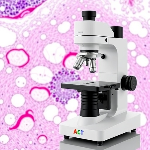
To showcase the versatility and effectiveness of the HySIL framework, the researchers engineered a modular device named SCOPE, which seamlessly augments existing light-sheet microscopy systems. This plug-and-play addition enables institutions to retrofit their current imaging setups to achieve best-in-class volumetric resolution affordably. Further pushing the envelope, the team developed a high-resolution variant dubbed Super-SCOPE, demonstrating the platform’s scalability and potential for refinement.
“By rethinking the role of immersion liquids from passive fillers to active optical elements, we have destroyed the historical trade-off between performance and accessibility in microscopy,” Professor Tomer stated. “Our HySIL-based systems achieve resolution comparable to the most expensive, oil-immersion setups but with a radically reduced footprint and cost, opening the door for use in diverse environments—from teaching laboratories to resource-limited clinical settings.”
These innovative optics have also been integrated into the compact, projector-based light-sheet microscope (pLSM) developed by the same group in 2024, now commercially distributed under the name SLICE. This commercialization trajectory promises broad dissemination of the technology, catalyzing next-generation research and diagnostics worldwide.
Collaborating with a rich network of academic partners, the team applied the pLSM-SCOPE system to image whole brains from mouse, salamander, and cavefish models, facilitating comprehensive neural circuit mapping essential for understanding brain function and evolution. They further extended applications to lab-grown miniature human brain organoids, advancing developmental biology and disease modeling. Notably, the technology was also employed in examining intact human cancer biopsies, heralding a paradigm shift in three-dimensional pathology and prognostication.
The HySIL framework’s broad compatibility means it can be adapted to other 3D imaging modalities beyond light-sheet microscopy, including confocal and two-photon systems, vastly expanding its utility. This adaptability positions HySIL as a foundational technology poised to accelerate tissue imaging research across multiple disciplines.
The impact of this technology extends beyond pure research. “By enabling scalable, high-resolution 3D imaging of tissues, we can fuel the emergence of advanced AI tools that revolutionize disease detection, grading, and outcome prediction,” Tomer emphasized. This capability is key for integrating large-scale tissue datasets into clinical workflows, promoting personalized medicine.
Industry partners have recognized the technology’s potential. Jack Glaser, CEO of MBF Bioscience and co-author of the study, expressed enthusiasm: “HySIL offers the rare combination of affordability and high performance, breaking down barriers for widespread adoption. Its engineering robustness and support infrastructure make it ideal for routine use in domains like neuroscience and cancer research.”
Historically, tissue analysis has depended heavily on thin, two-dimensional slices placed on glass slides, a method that overlooks critical spatial context inherent to complex biological systems. By empowering researchers and clinicians with accessible 3D imaging tools, technologies like pLSM-SCOPE stand to redefine diagnostic standards and biological inquiry.
Pathology expert Hanina Hibshoosh, who co-authored the paper, underscored this advancement: “Visualizing tissue architecture in three dimensions reveals essential patterns and anomalies missed in traditional cross-sectional views. Affordable instruments that democratize such capabilities will become indispensable as AI-driven analyses expand.”
Supported by the National Institutes of Health and several academic institutions, this work highlights the fruitful intersection of optics innovation, computational imaging, and biomedical research. Columbia University has also filed patents to protect and facilitate the deployment of HySIL and its associated technologies. Importantly, the collaboration with MBF Bioscience ensures practical pathways from laboratory innovation to commercial availability.
Many individuals contributed to this milestone, including faculty members, postdoctoral fellows, doctoral candidates, and research associates, reflecting a deeply interdisciplinary effort critical for solving complex scientific challenges.
Ultimately, the HySIL concept represents a paradigm shift in microscope optics. By enabling superior imaging at reduced cost and complexity, it democratizes access to detailed 3D tissue data, fueling discoveries and clinical insight. As biological and medical sciences increasingly hinge on comprehensive spatial information, technologies like HySIL will be instrumental in charting the future of research and healthcare.
Subject of Research: Human tissue samples
Article Title: Hybrid solid−liquid optics enable scalable, high-resolution light-sheet microscopy across diverse immersion media
News Publication Date: 9-Jun-2026
Web References: https://doi.org/10.1038/s41587-026-03172-7
Image Credits: Imaged with SCOPE, a device developed by Professor Raju Tomer and colleagues at Columbia University
Keywords
3D tissue imaging, HySIL, light-sheet microscopy, SCOPE, Super-SCOPE, high-resolution optics, breast tissue imaging, neural circuit mapping, pathology, AI diagnostics, optical lenses, microscope innovation
Tags: 3D tissue imaging technologyadvanced microscopy designaffordable biological imaging toolsAI diagnostic models in microscopybiological research microscopy advancementsbrain tissue imaging innovationscancer biopsy 3D visualizationhigh-resolution volumetric microscopyimaging depth versus sharpness trade-offnon-oil immersion microscopy benefitsovercoming optical lens limitationsscalable microscope technology

