A groundbreaking advancement in the realm of neuroimaging has emerged, unveiling a sophisticated and quantitative method to evaluate synaptic density in the spinal cord of multiple sclerosis (MS) patients. This novel positron emission tomography (PET) imaging technique employs the synaptic vesicle glycoprotein 2A (SV2A) tracer, offering unprecedented insights into the intricate neural connections that are compromised in the course of MS. Synapses, the microscopic junctions facilitating communication between neurons, are critical to maintaining the brain and spinal cord’s functional integrity. The capacity to measure synaptic loss in vivo represents a transformative stride toward personalizing disease monitoring and assessing therapeutic efficacy in MS, a disease renowned for its complex neuroinflammatory and neurodegenerative pathology.
Multiple sclerosis, a chronic autoimmune disorder, primarily disrupts the myelin sheath encasing nerve fibers in the central nervous system. However, beyond demyelination lies a subtler yet devastating form of neural injury—the degeneration of synapses. Until recently, non-invasive quantification of synaptic density, especially within the spinal cord, was elusive due to technical and anatomical challenges. The spinal cord is a critical nexus for motor and sensory pathways and serves as a primary locus for early neuroinflammatory processes in MS. Consequently, the ability to quantify synaptic loss at this site could elucidate mechanisms underlying symptomatology and disease progression that have been difficult to trace by conventional imaging modalities.
In this pioneering study, researchers utilized the radiotracer [^18F]SynVesT-1, a ligand selective for SV2A, to conduct PET imaging in mice with experimental autoimmune encephalomyelitis (EAE), a well-established animal model of MS. The EAE model replicates many features of human MS, including neuroinflammation and demyelination. By delineating regions of interest within the spinal cord and quantifying tracer uptake—expressed as total volume of distribution (VT)—the investigators demonstrated significant reductions in synaptic density in affected animals compared to healthy controls. These findings were corroborated by ex vivo binding assays, confirming that SV2A PET accurately reflects synaptic alterations in this disease context.
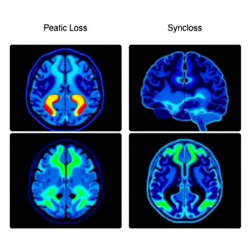
Building on the preclinical evidence, the researchers translated their approach to human subjects using [^11C]UCB-J PET imaging, another SV2A-specific radioligand with well-characterized binding properties in humans. They conducted imaging on a cohort comprising six MS patients and an equivalent number of healthy controls, meticulously generating VT maps to assess synaptic density across diverse brain and spinal cord regions. The patients exhibited a notable 16.4% reduction in cortical and subcortical synaptic density relative to controls, underscoring the widespread synaptic pathology characteristic of MS. Crucially, similar patterns of synaptic loss emerged in the spinal cord, reinforcing the translational relevance of the animal model findings.
This comprehensive cross-species analysis has validated SV2A PET as a sensitive and quantitative biomarker for synaptic pathology in multiple sclerosis. The ability to noninvasively measure synaptic density alterations in vivo opens a new chapter for both clinical research and patient care. Clinicians could leverage this technology for real-time monitoring of disease progression, moving beyond traditional imaging markers that primarily highlight gross structural damage and demyelination. Furthermore, SV2A PET promises to serve as an invaluable tool for evaluating novel therapeutics aimed at synaptic preservation and restoration, which are increasingly recognized as vital targets for ameliorating MS symptoms and disability.
The technical sophistication of SV2A PET stems from its molecular specificity and capacity to capture dynamic synaptic changes at cellular resolution. Synaptic vesicle glycoprotein 2A, ubiquitously expressed in presynaptic terminals, serves as a proxy for total synaptic density. Radiolabeled ligands such as [^18F]SynVesT-1 and [^11C]UCB-J exhibit high affinity and favorable kinetic profiles, enabling precise quantification of synaptic loss. Unlike conventional MRI techniques that visualize macroscopic lesions or atrophy, SV2A PET uncovers subtle synaptic alterations, revealing the extent of neurodegeneration that correlates with clinical manifestations and cognitive decline in MS.
The pioneering work described here also provides important insights into the spatial distribution of synaptic pathology. Both preclinical and human data indicate that synaptic loss is not restricted to white matter lesions but is pervasive across cortical, subcortical, and spinal cord regions. This widespread involvement may explain the heterogeneity of symptoms experienced by patients, ranging from motor deficits and sensory disturbances to fatigue and cognitive impairment. Consequently, SV2A PET imaging offers a promising avenue for dissecting the complex pathophysiology of MS and tailoring individualized therapeutic strategies based on synaptic integrity.
Although this imaging approach is currently limited to select research settings and clinical trials due to the need for specialized radiochemistry and scanner infrastructure, its implications for the future of MS management are profound. Larger, multicenter studies are warranted to validate these initial findings and establish standardized protocols for clinical deployment. If validated, SV2A PET could become an integral part of precision medicine initiatives, enabling neurologists to detect synaptic degeneration earlier, monitor response to emerging neuroprotective drugs, and ultimately improve patient outcomes.
The ongoing evolution of neuroimaging technologies encodes a shift towards elucidating the functional and molecular substrates of neurological diseases. By integrating SV2A PET with complementary imaging modalities and clinical assessments, researchers and clinicians can gain a holistic understanding of disease mechanisms in multiple sclerosis. This multidimensional approach promises to accelerate drug development pipelines and enhance the granularity of patient monitoring, addressing an unmet need in the care of a condition marked by unpredictable progression and variable treatment responses.
From a mechanistic perspective, the findings underscore the critical role of synaptic integrity in maintaining neurological function and the devastating impact of synaptic loss in MS. Therapeutic strategies focused solely on remyelination may be insufficient to halt clinical deterioration if synaptic connections continue to degenerate. Therefore, SV2A PET not only aids in diagnosis and monitoring but could also facilitate novel interventions targeting synaptic repair and synaptogenesis, heralding a paradigm shift in neurotherapeutics.
In summary, the innovative application of SV2A PET imaging to both animal models and human patients reveals synaptic density loss as a pervasive and quantifiable hallmark of multiple sclerosis pathology. This technological breakthrough enables in vivo visualization of neural circuit damage, providing crucial insights that bridge preclinical and clinical neuroscience. As this field advances, SV2A PET stands poised to revolutionize how neurologists understand, track, and ultimately treat the synaptic underpinnings of MS, offering hope for more precise, effective, and personalized healthcare interventions.
Subject of Research: Synaptic density measurement and imaging in multiple sclerosis using SV2A PET.
Article Title: Tracking Synaptic Density Loss in the Spinal Cord of Experimental Autoimmune Encephalomyelitis Mice and Pilot Evaluation of SV2A PET in Patients with Multiple Sclerosis.
News Publication Date: 2026 (presented at the 2026 Society of Nuclear Medicine and Molecular Imaging Annual Meeting).
Web References:
https://www.snmmi.org/
https://www.xcdsystem.com/snmmi/program/UtDKfSi/index.cfm?pgid=3058&sid=53922&mobileappid=5392200000
References:
Chia PHJ, Le H, Tong J, Alijaniaram M, Vasdev N, Zheng C; Toyonaga T, Chen M-K. Journal of Nuclear Medicine.
Image Credits: Courtesy of Society of Nuclear Medicine and Molecular Imaging (SNMMI).
Keywords: Multiple sclerosis, synaptic density, SV2A PET, neuroimaging, experimental autoimmune encephalomyelitis, positron emission tomography, [^18F]SynVesT-1, [^11C]UCB-J, spinal cord imaging, neurodegeneration, personalized medicine, molecular imaging.
Tags: advances in non-invasive neuroimaginghuman PET imaging of synaptic lossmolecular imaging biomarkers for MS progressionneurodegenerative mechanisms in MS spinal cordneuroinflammation and synaptic degeneration in MSpersonalized monitoring of multiple sclerosisPET imaging for synaptic density in multiple sclerosispreclinical PET studies in multiple sclerosisspinal cord synaptic loss quantificationsynaptic pathology in autoimmune neurodegenerative diseasessynaptic vesicle glycoprotein 2A tracer in neuroimaging

