In a groundbreaking study set to redefine the landscape of neurodegenerative disease diagnosis, researchers have harnessed the power of advanced machine learning and ultra-high-field magnetic resonance imaging (MRI) to enhance the early identification of Parkinson’s disease. Leveraging a support vector machine (SVM) model driven by complex, multidimensional data obtained from 7-Tesla structural MRI scans, the team proposes a transformative diagnostic approach that promises unprecedented precision. This novel technique represents a significant leap forward in the detection and understanding of Parkinson’s disease pathology, potentially enabling clinicians to intervene earlier and with greater confidence.
Traditionally, Parkinson’s disease diagnosis has relied heavily on clinical evaluation of motor symptoms and ancillary tests that often detect the disease at relatively late stages. This delay hampers the effectiveness of therapeutic strategies aimed at slowing disease progression. However, the integration of neuroimaging biomarkers with machine learning algorithms heralds a new era wherein subclinical changes in brain structures can be detected far earlier. The study under discussion exploits the superior spatial resolution and tissue contrast afforded by 7-Tesla MRI, which surpasses the capabilities of conventional 1.5T and 3T scans, to capture subtle abnormalities within the brain’s architecture.
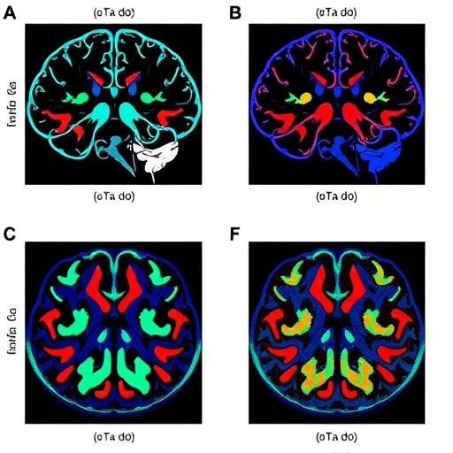
At its core, the team employed support vector machines, a type of supervised machine learning model known for its excellent handling of high-dimensional data and robust classification performance. By training the SVM with structural MRI features extracted from both patients diagnosed with Parkinson’s disease and healthy controls, the researchers constructed a classification algorithm capable of discerning complex patterns of neuroanatomical change characteristic of the disease. Importantly, the multidimensional nature of the input data included volumetric measurements, cortical thickness, and microstructural integrity parameters, providing a comprehensive anatomical profile.
The utility of 7-Tesla MRI in this framework cannot be overstated. The ultra-high field strength enhances signal-to-noise ratio, allowing for finer-grained visualization of brain regions critically implicated in Parkinson’s pathophysiology, such as the substantia nigra, basal ganglia, and associated white matter tracts. These regions often exhibit subtle degeneration not easily captured through lower-field imaging. Using specialized imaging sequences, the researchers acquired structural data that underpin the SVM’s capacity to detect disease-related alterations with remarkable sensitivity.
In practical terms, the study involved scanning a sizable cohort consisting of both early-stage Parkinson’s disease patients and age-matched healthy individuals. Structural MRIs were preprocessed to extract a battery of quantitative biomarkers representing brain morphology and integrity. These imaging features were then inputted into the SVM model, which underwent rigorous cross-validation to optimize its classification thresholds and avoid overfitting. The results indicated that the SVM-driven approach achieved superior accuracy compared to existing diagnostic methods, significantly reducing false negatives and false positives.
Beyond diagnostic accuracy, the machine learning model provides an interpretable framework to understand which structural changes most strongly predict disease presence. Feature importance analysis revealed that specific volumetric reductions in the substantia nigra pars compacta, alterations in cortical thickness of frontal and temporal regions, and disruptions in white matter microstructure, measured by advanced diffusion metrics, emerged as key indicators. These findings enrich the neurobiological understanding of Parkinson’s disease and may guide future biomarker development.
The study’s implications extend beyond diagnosis, potentially informing patient stratification for clinical trials and individualized treatment planning. By pinpointing patients in their prodromal or early clinical stages, therapeutic interventions can be tailored before irreversible neuronal loss occurs. Moreover, the methodology paves the way for longitudinal tracking of disease progression through imaging biomarkers, enabling more precise monitoring of treatment efficacy.
Integration of artificial intelligence with high-resolution imaging also addresses the challenge of diagnostic variability inherent in clinical assessments. Subjectivity and inter-rater differences often complicate Parkinson’s diagnosis, but a standardized, algorithm-driven process introduces objectivity and scalability. As healthcare systems increasingly adopt digital tools, this combined approach could be embedded into routine neurology workflows, facilitating wider access to early and accurate diagnosis.
Crucially, the multidisciplinary collaboration between neuroimaging specialists, machine learning experts, and clinical neurologists underlines the importance of cross-sector innovation in tackling complex brain disorders. The researchers emphasize that the success of the SVM-driven diagnostic model is attributable not only to advanced computational techniques but also to high-quality imaging data and careful clinical phenotyping.
While promising, the study acknowledges several limitations and areas for future research. Larger, multicenter cohorts are necessary to validate the model’s generalizability across diverse populations. Additionally, combining structural MRI with other modalities such as functional MRI, positron emission tomography (PET), or cerebrospinal fluid biomarkers could enhance diagnostic comprehensiveness. Investigations into automated workflows for MRI acquisition and processing would further improve clinical adoption.
The ethical considerations around AI-based diagnostics are also discussed. Transparency regarding algorithm decision-making, data privacy, and patient consent remain paramount. The researchers advocate for robust governance frameworks to ensure responsible integration of AI tools in clinical practice, supporting equitable and beneficial outcomes for patients.
In summary, this innovative study demonstrates that the fusion of support vector machine algorithms with 7-Tesla multidimensional structural MRI data represents a powerful tool for the early identification of Parkinson’s disease. By moving beyond symptom-based diagnosis toward objective, imaging-derived biomarkers, this approach holds promise for revolutionizing patient care and accelerating therapeutic advancements. As the technology matures, it may also be adapted to other neurodegenerative disorders, broadening its impact.
The convergence of ultra-high-field neuroimaging and artificial intelligence exemplifies the future of precision medicine in neurology. This landmark research not only advances scientific understanding but also offers hope to millions affected by Parkinson’s disease worldwide, highlighting a path toward earlier diagnosis, improved intervention strategies, and ultimately, better quality of life.
Subject of Research:
Article Title:
Article References:
Xiong, Y., Li, Z., Yang, M. et al. Support vector machine-driven Parkinson’s disease identification: a 7-Tesla multidimensional structural MRI approach. npj Parkinsons Dis. (2026). https://doi.org/10.1038/s41531-026-01370-3
Image Credits: AI Generated
Tags: 7-Tesla MRI for Parkinson’s detectionadvanced neuroimaging biomarkersearly detection of neurodegenerative diseasesearly intervention in Parkinson’shigh-resolution structural MRIimproving Parkinson’s diagnostic accuracymachine learning for Parkinson’s diagnosismultidimensional data analysis in MRIParkinson’s disease pathology imagingsupport vector machine in neuroimagingSVM algorithm in medical imagingultra-high-field MRI brain scans

