A groundbreaking study emerging from the University of Nottingham has unveiled the elusive behavior of the human placenta during pregnancy, revealing that it contracts independently of the surrounding uterine wall in a significant majority of healthy pregnancies. This discovery, documented through advanced magnetic resonance imaging (MRI) techniques, challenges long-held assumptions about placental dynamics and opens new avenues for monitoring fetal health with unprecedented precision.
The placenta, often described as the lifeline between mother and fetus, plays a critical role in nutrient and oxygen exchange, waste removal, and hormonal regulation. Despite its importance, clinical monitoring of placental function has historically relied on indirect markers such as fetal growth patterns and movement, lacking direct observation of the organ’s own activity. Early MRI studies hinted at placental contractions separate from uterine contractions, but the characteristics and differentiation of these contractions remained ambiguous until now.
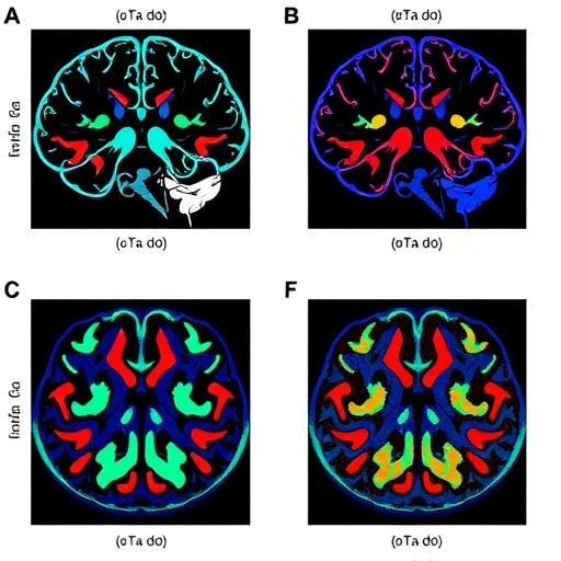
In this latest research, a cohort of 36 healthy pregnant women between the gestational ages of 29 and 42 weeks underwent MRI scanning sessions that lasted anywhere from 15 to 32 minutes. During these sessions, high-resolution axial images of the uterus were captured at multiple time points, allowing an unprecedented real-time glimpse into placental and uterine dynamics. To handle the complex datasets generated, researchers employed automated neural networks to analyze morphological changes within the placenta and uterine tissues, focusing on volumetric, areal, and shape alterations.
One of the most striking findings was that placental contractions were identified in around 60% of the pregnancies studied, occurring at a frequency averaging about two per hour. These contractions lasted longer on average than typical uterine contractions, with a median duration of approximately 2.4 minutes. Unlike uterine contractions that predominantly influence the uterine wall’s structure, placental contractions were uniquely associated with notable shape changes of the placenta itself, particularly in its sphericity—a measure of how sphere-like the organ becomes during contraction.
The study also mapped biochemical changes associated with these contractions through an MRI parameter known as R2, which correlates with levels of deoxygenated blood. Both placental and uterine contractions caused an increase in placental R2 values, indicating transient rises in deoxygenated blood during contractions. This insight hints at dynamic blood flow and oxygenation changes that may be critical to maintaining a healthy placental environment and, consequently, fetal well-being.
Employing sophisticated segmentation algorithms, the team was able to distinguish placental contractions from uterine contractions automatically by analyzing specific shape metrics, notably the degree of sphericity. This breakthrough paves the way for real-time, non-invasive monitoring tools capable of providing clinicians with direct, objective measurements of placental function, potentially improving the early detection of pregnancy complications.
Despite its pioneering nature, the study acknowledges certain limitations. The sample size was relatively modest, and the MRI scan durations, while sufficient for initial characterization, were short relative to the entire pregnancy timeline, leaving open questions about the frequency and behavior of these contractions over longer periods. Not all participants exhibited both placental and uterine contractions during the imaging window, highlighting the need for extended monitoring in future research.
The implications of these findings are substantial, particularly for understanding conditions linked with placental insufficiency, such as fetal growth restriction and pre-eclampsia. Dr. Louise Dewick, lead author and researcher at the University of Nottingham’s School of Medicine, emphasized the critical nature of this discovery: “By characterizing placental contractions in vivo, we have created a foundation for future studies aimed at unraveling complex placental pathologies, which could ultimately transform prenatal care.”
The technological advancements underpinning this research also signal a new era in obstetric imaging. Professor Penny Gowland, from the School of Physics and Astronomy, highlighted how enhanced MRI methods allow unprecedented visualization of the gravid uterus, demonstrating that the placenta is a highly dynamic organ, not merely a passive interface. These bio-imaging breakthroughs could lead to routine clinical applications for identifying placental distress well before symptoms manifest.
Multidisciplinary collaboration was central to the success of this groundbreaking work. Amy Turnball, a member of the research team, remarked on the rewarding experience of working across fields, bringing together clinicians, physicists, and data scientists to unravel this complex biological phenomenon. The collective expertise enabled the deployment of neural networks for image analysis, a technique rapidly gaining prominence in medical imaging for its precision and efficiency.
The findings accentuate that placental contractions are a physiological phenomenon previously underrecognized in clinical practice. The recognition that the placenta itself actively contracts, potentially influencing uteroplacental blood flow and oxygen availability, introduces a novel dimension to our understanding of the maternal-fetal interface. This dynamic perspective could catalyze innovations for monitoring fetal health and managing pregnancies at risk due to placental dysfunction.
Looking ahead, future investigations are needed to evaluate how placental contraction patterns may vary in complicated pregnancies. Longitudinal studies with larger participant numbers and extended monitoring durations will be necessary to establish normative contraction profiles and identify deviations indicative of pathology. Such research could ultimately influence guidelines for prenatal care, particularly in high-risk populations.
In sum, this study not only uncovers a vital physiological function of the placenta but also demonstrates the power of state-of-the-art imaging and artificial intelligence to push the boundaries of prenatal diagnostics. As the placenta steps into the spotlight as an active participant in pregnancy regulation, clinicians and researchers are poised to unlock new strategies to safeguard maternal and fetal health, improving outcomes worldwide.
Subject of Research: People
Article Title: Placental contractions in uncomplicated pregnancies
News Publication Date: April 29, 2026
Web References:
Article DOI: http://dx.doi.org/10.1371/journal.pone.0344388
Wellcome Leap In Utero Programme: https://wellcomeleap.org/inutero/program/
References:
Dewick L, Turnbull A, Walker K, Jones N, Hutchinson G, Bradley C, et al. (2026) Placental contractions in uncomplicated pregnancies. PLoS One 21(4): e0344388.
Image Credits: Dewick et al., 2026, PLOS One, CC-BY 4.0
Keywords: Placental contractions, MRI, pregnancy, uterine wall, fetal health, placental monitoring, deoxygenated blood, neural network, placental shape, placental dysfunction, fetal growth restriction, pre-eclampsia
Tags: advanced MRI techniques for placentafetal health monitoring innovationsindependent placental activity MRInon-invasive fetal health diagnosticsnutrient and oxygen exchange in placentaplacental and uterine wall dynamicsplacental contraction frequency researchplacental contractions in healthy pregnanciesplacental function assessment methodspregnancy gestational age MRI scansreal-time placental imagingUniversity of Nottingham pregnancy study