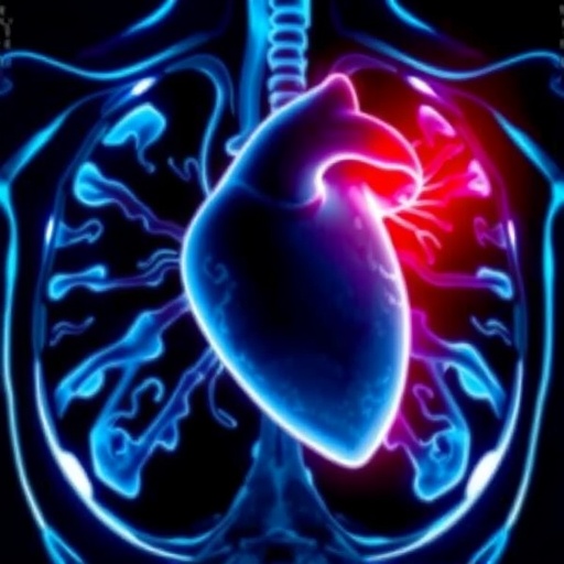
A routine cardiac computed tomography (CT) scan, long utilized as a non-invasive method to assess coronary artery blockages, is undergoing a revolutionary transformation. Researchers at Kumamoto University have unveiled a method that leverages this common imaging technique to expose hidden heart muscle damage, offering an unprecedented prognostic insight into future cardiac events. This evolution of cardiac CT not only enhances diagnostic precision but also empowers clinicians to predict and preempt heart failure and mortality with significantly higher accuracy.
Traditionally, cardiac CT scans have served the singular purpose of evaluating coronary artery conditions, primarily focusing on detecting stenoses and plaques that may precipitate myocardial infarctions. However, the conventional application overlooks subtle yet clinically significant alterations within the myocardium itself. The groundbreaking work by Professors Yasuhiro Izumiya and Kenichi Tsujita’s team expands the utility of CT imaging by incorporating a delayed imaging phase designed to identify molecular and structural cardiac tissue changes indicative of damage.
This approach hinges on two powerful markers derived from delayed-phase CT imaging: Late Iodine Enhancement (LIE) and Extracellular Volume (ECV). LIE pinpoints localized myocardial scarring by detecting iodine accumulation in fibrotic tissue regions, similar in principle to late gadolinium enhancement observed in cardiac MRI but adapted for CT technology. Conversely, ECV measures the fraction of extracellular space, serving as a surrogate for diffuse interstitial fibrosis and subtle myocardial injury that conventional CT or other imaging modalities might miss.
The strength of combining these markers lies in their complementary nature. LIE captures distinct scars, often the sequelae of previous infarcts or focal injury, while ECV captures more diffuse, less conspicuous alterations in myocardial integrity. Together, they present a holistic depiction of cardiac tissue health that goes beyond anatomical assessment, venturing into the realm of pathophysiology with non-invasive tomography.
In the observational study encompassing 1,207 patients with an average follow-up duration of 26 months, the presence of abnormalities in both LIE and ECV served as a robust harbinger of adverse clinical outcomes. Patients exhibiting elevated readings on both fronts demonstrated a notably higher incidence of unplanned hospitalizations due to cardiac causes and increased mortality rates, underscoring the dual markers’ prognostic power for long-term cardiovascular risk stratification.
These findings challenge the traditional paradigm wherein coronary artery imaging alone dictates risk assessment and guide therapeutic decisions. The novel insight that subtle myocardial damage, elusive to standard imaging, possesses independent predictive value could reshape clinical pathways. It suggests that cardiac CT can transcend its conventional role to simultaneously evaluate coronary anatomy and myocardial condition in a single session.
The implications for clinical practice are extensive. The modified CT protocol is faster and more accessible than cardiac magnetic resonance imaging (MRI), which is often considered the gold standard for myocardial tissue characterization but is costly, less available, and contraindicated in certain patient populations. Consequently, this CT enhancement grants broader reach, especially in environments with limited MRI access, democratizing advanced cardiac diagnostics.
Additionally, earlier detection of occult myocardial damage enables timely interventions which can alter disease trajectories. Physicians can stratify patients more accurately and tailor treatments to mitigate progression to heart failure, potentially reducing mortality and healthcare burden. This aligns perfectly with the evolving ethos of precision medicine, where individualized risk prediction and management improve outcomes.
Technological advancements in CT hardware and software have been critical enablers of this breakthrough. Improvements in detector sensitivity, image resolution, and post-processing algorithms allow the differentiation of iodine kinetics and tissue characteristics with remarkable clarity. These technical refinements have transformed a conventional imaging modality into a dual-purpose tool with diagnostic and prognostic capabilities.
Moreover, the radiation exposure concerns traditionally associated with CT scanning are being mitigated by emerging low-dose protocols and enhanced reconstruction techniques. These advances ensure that the benefits of extended cardiac tissue evaluation do not come at the expense of patient safety, preserving the risk-benefit balance in diagnostic imaging.
The researchers emphasize that the synergy of LIE and ECV offers a comprehensive perspective on cardiac pathology that isolated markers cannot achieve. This nuanced detection strategy holds promise not only for patients at risk for ischemic heart disease but also for those with cardiomyopathies, myocarditis, and other myocardial disorders where fibrosis and extracellular remodeling play pivotal roles.
As this technique gains traction, it is poised to become a cornerstone in cardiac imaging, redefining how clinicians conceptualize and manage heart disease. The integration of structural, functional, and pathological insights within a single scan heralds a new era in cardiovascular medicine, where obscured dangers are illuminated before advancing to the stage of clinical crisis.
In conclusion, the addition of a delayed phase to routine cardiac CT scanning represents a significant stride toward comprehensive cardiac care. By uncovering hidden myocardial injury through the combined assessment of LIE and ECV, this method offers an advanced, non-invasive, and practical means to predict patient outcomes better and personalize treatment strategies. This promising paradigm shift holds the potential to save lives and reduce the burden of cardiovascular diseases globally.
Subject of Research: Not applicable
Article Title: Does adding a delayed phase to cardiac computed tomography for coronary artery evaluation have prognostic value?
News Publication Date: 22-Jan-2026
Web References: http://dx.doi.org/10.1093/ehjci/jeag018
References: Oguni et al., European Heart Journal Cardiovascular Imaging (2026), licensed under Creative Commons Attribution 4.0 International (CC BY 4.0).
Image Credits: Oguni et al.
Keywords: Heart disease, Coronary artery disease, Cardiovascular disorders, Medical imaging, Tomography, Clinical medicine
Tags: cardiac CT scan advancementscoronary artery blockage imagingdelayed-phase CT imaging techniquesenhanced cardiac diagnostic precisionExtracellular Volume measurement in myocardiumKumamoto University cardiac researchLate Iodine Enhancement in cardiac CTmolecular cardiac tissue analysismyocardial scarring detection methodsnon-invasive heart muscle damage detectionpredicting heart failure with CT scansprognostic cardiac risk assessment

