In a groundbreaking convergence of neuroimaging and molecular genetics, researchers have unveiled compelling insights into the spatial relationship between gene expression and brain atrophy patterns characteristic of dementia with Lewy bodies (DLB). This pioneering study leverages advanced imaging transcriptomics, offering an unprecedented window into the molecular underpinnings of neurodegeneration in this enigmatic disorder. The findings promise to reshape our understanding of DLB’s pathophysiology and open up new avenues for targeted therapeutic interventions.
Dementia with Lewy bodies is a complex neurodegenerative disease marked by progressive cognitive decline, fluctuating attention, visual hallucinations, and parkinsonian motor symptoms. Despite advances in clinical diagnosis and symptomatic treatments, the molecular mechanisms driving selective vulnerability and regional brain deterioration remain insufficiently understood. The integration of high-resolution brain imaging with regional gene expression profiling presents a transformative approach to disentangle these mechanisms by linking anatomical changes to underlying molecular dysfunctions.
In this study, spearheaded by Habich, Baumann, Schwarz, and their colleagues, researchers employed imaging transcriptomics, a cutting-edge methodological framework that combines neuroimaging data with transcriptome maps. This approach helps pinpoint the locational alignment of gene expression patterns with observed cortical thinning and subcortical atrophy in DLB patients. By cross-referencing large-scale transcriptomic databases with structural MRI scans, the team delineated gene-brain atrophy correlations that shed light on region-specific susceptibilities.
One of the pivotal discoveries demonstrated that distinct clusters of genes involved in synaptic function, lysosomal activity, and proteostasis align spatially with regions exhibiting significant atrophic changes. Notably, genes implicated in alpha-synuclein metabolism—a hallmark protein aggregate in DLB—showed enriched expression within atrophy-prone areas. This reinforces the pathological relevance of alpha-synuclein’s misprocessing and accumulation in shaping neurodegenerative trajectories.
Moreover, the study revealed that brain regions with pronounced atrophy were enriched for gene sets regulating immune responses and neuroinflammation. These findings resonate with emerging literature positing neuroinflammation as a critical driver of neurodegeneration, suggesting a bidirectional interplay between immune activation and neuronal loss in the DLB brain. Importantly, this spatial-genetic linkage provides a tangible molecular signature that could be harnessed for biomarker development and personalized medicine.
A highlight of the research involves the identification of regionally selective vulnerability that could explain the clinical heterogeneity observed among DLB patients. By mapping transcriptomic landscapes onto atrophic patterns, investigators uncovered genetic profiles unique to vulnerable subcortical nuclei and cortical regions governing cognition and motor control. This granular insight paves the way for tailored interventions targeting region-specific molecular pathways.
Technological breakthroughs in multi-modal data integration underpinned the success of the study. High-resolution magnetic resonance imaging furnished precise quantifications of gray matter volume reductions, while publicly accessible brain transcriptome atlases enabled comprehensive gene expression mapping. Sophisticated bioinformatics pipelines coupled these datasets, allowing robust statistical inference while mitigating confounding factors such as age-related changes and comorbidities.
The implications extend far beyond academic understanding, as this approach could refine patient stratification in clinical trials. By incorporating individual genetic expression patterns correlated with neuroanatomical degeneration, clinicians may predict disease progression more accurately and optimize therapeutic strategies. This biomarker-driven framework marks a pivotal shift from symptomatic to mechanism-targeted treatments in DLB.
Furthermore, the findings challenge the traditional notion that neurodegeneration propagates uniformly across brain tissue. Instead, the study underscores that gene expression heterogeneity contributes decisively to which neuronal populations succumb or resist pathological insult. Recognizing this, future research might explore gene editing or modulation techniques in spatially defined circuits as potential disease-modifying therapies.
Beyond neurodegeneration, the imaging transcriptomic methodology showcased here holds promise for a spectrum of neurological disorders characterized by region-specific brain changes. Alzheimer’s disease, frontotemporal lobar degeneration, and even psychiatric illnesses such as schizophrenia could benefit from similar integrative analyses, potentially unraveling novel pathogenic pathways and therapeutic targets.
Despite its transformative potential, the research acknowledges limitations, including reliance on postmortem gene expression data from healthy brains, which may not fully recapitulate disease-altered transcriptional dynamics. Longitudinal studies incorporating patient-specific transcriptomics combined with in vivo neuroimaging could address these gaps and refine causative inferences.
In sum, this landmark imaging transcriptomics study presents a compelling framework that bridges the molecular and structural landscapes of dementia with Lewy bodies. By delineating the genetic signatures underpinning regional brain atrophy, the research illuminates the intricate molecular choreography driving neurodegeneration and opens promising therapeutic horizons. As technological capabilities evolve, integrating genotype, phenotype, and neuroimaging data stands to revolutionize our battle against complex brain disorders.
The study’s insights signal an exciting era in neuroscience where dissecting the molecular geography of the brain translates directly into clinical impact. With dementia prevalence rising globally and therapeutic progress lagging, such innovative interdisciplinary efforts provide a beacon of hope for patients, caregivers, and clinicians alike. The intersection of gene expression patterns and neuroanatomy charts a path toward precision neurology that could transform prognostic models and ultimately, patient care.
As further exploration into transcriptomic signatures within neurodegeneration accelerates, the anticipation builds for novel biomarker panels and gene-targeted therapeutics inspired by this study. The nexus of molecular biology and brain imaging not only decodes disease mechanisms but also personalizes intervention strategies at an individual brain region level. This paradigm shift could redefine diagnostics and treatments, heralding a future where dementia is more predictable, treatable, and one day possibly preventable.
The lasting contribution of this research lies in its elegant demonstration of how complex diseases like DLB emerge from the intricate interplay between genes and brain structure. It exemplifies how harnessing big data and computational neuroscience can yield profound biological insights previously inaccessible. The lessons learned here will undoubtedly spur similar integrative endeavors, accelerating discoveries across neurological and psychiatric landscapes.
Above all, this study mobilizes a fresh perspective on DLB—conceiving the brain not merely as a uniform organ vulnerable to global decline but as a mosaic of molecularly distinct areas whose selective degeneration orchestrates clinical symptomatology. This nuanced understanding empowers researchers and clinicians to envision precision interventions tailored to each patient’s unique molecular-neuroanatomical signature, fundamentally transforming neurology’s approach to neurodegenerative disease.
Subject of Research: Regional gene expression and brain atrophy in dementia with Lewy bodies.
Article Title: Regional gene expression and brain atrophy in dementia with Lewy bodies: an imaging transcriptomics study.
Article References:
Habich, A., Baumann, J.M., Schwarz, C.G. et al. Regional gene expression and brain atrophy in dementia with Lewy bodies: an imaging transcriptomics study. npj Parkinsons Dis. 12, 96 (2026). https://doi.org/10.1038/s41531-026-01355-2
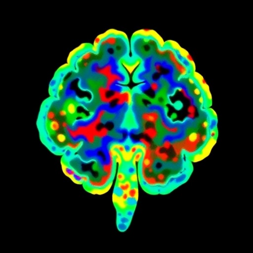
Image Credits: AI Generated
DOI: https://doi.org/10.1038/s41531-026-01355-2
Tags: brain atrophy patterns in DLBcortical thinning in Lewy body dementiaimaging transcriptomics in neurodegenerationLewy body dementia gene expressionmolecular mechanisms of dementianeuroimaging and transcriptome integrationParkinsonian symptoms molecular basispathophysiology of Lewy body dementiaspatial gene expression brain mappingsubcortical atrophy gene mappingtargeted therapies for DLBtranscriptomic profiling in neurodegenerative diseases


