In a groundbreaking advancement that promises to revolutionize the monitoring of stem cell therapies in cardiac medicine, researchers at the Institute of Biomedical Engineering at the University of Toronto have pioneered a novel magnetic resonance imaging (MRI) technique known as “bright ferritin MRI.” This innovative approach enables the prolonged tracking of transplanted human pluripotent stem cell-derived cardiomyocytes in rat hearts for periods extending up to eight weeks. Such a capability marks a significant leap forward in regenerative cardiology, addressing a persistent challenge in the visualization of therapeutic cell survival and integration within host tissues.
Stem cell-based therapies offer unprecedented potential to repair heart damage caused by myocardial infarction, primarily through the regeneration of functional cardiac muscle cells. Human pluripotent stem cell-derived cardiomyocytes, specifically, possess the ability to engraft and electrically couple with native myocardium, thereby restoring contractile function. However, a critical barrier to clinical translation has been the limited survival of transplanted cells, compounded by the inability to robustly monitor these cells over extended durations in vivo. Traditional imaging modalities have fallen short, either due to incompatibility with larger animal models or the transient nature and ambiguity of labeling signals that degrade as cells proliferate or engage the host immune response.
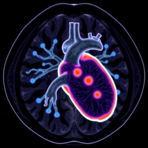
Addressing these limitations, Professor Hai-Ling Margaret Cheng and her team engineered a state-of-the-art imaging platform integrating the overexpression of ferritin, an intracellular iron-storage protein, within human pluripotent stem cells. By genetically modifying these cells to upregulate ferritin, the researchers endowed the cardiomyocytes with an intrinsic MRI contrast capability. This molecular engineering ensures that the iron sequestered by ferritin generates a distinct and enhanced signal under MRI, especially when augmented with manganese chloride administration, which induces a bright contrast, thus enabling three-dimensional spatial mapping of the surviving transplanted cells.
Critically, extensive in vitro characterization of these ferritin-overexpressing cardiomyocytes confirmed preservation of essential cellular phenotypes including normal architecture, contractile protein expression, and electrophysiological properties. Such validation underscores that the genetic modification does not compromise the fundamental cardiac functions requisite for therapeutic efficacy. Subsequently, the team transplanted the engineered cells into the left ventricular myocardium of immunodeficient rat models, both healthy and with induced cardiac injury, to emulate clinically relevant conditions.
Utilizing high-resolution MRI scanners, the researchers longitudinally tracked these cells over an eight-week period, a duration that surpasses typical tracking intervals recorded in previous studies. The administration of manganese chloride played a pivotal role in amplifying the ferritin-related MRI signal, facilitating the precise localization and persistence mapping of viable transplanted cells in living subjects. Post-mortem histological analyses corroborated the imaging data, confirming that the bright signals corresponded to genuine cell survival rather than artifacts or nonspecific uptake. Additionally, echocardiographic evaluation demonstrated that manganese treatment did not adversely affect cardiac function, affirming the safety profile of this imaging adjunct.
This technique answers a long-standing need in regenerative medicine by enabling clinicians and researchers to non-invasively visualize therapeutic cell engraftment with high spatial and temporal resolution. The ability to discern not only the presence but also the precise anatomical distribution of surviving stem cell-derived cardiomyocytes offers invaluable insights into mechanisms of cell survival, migration, and integration within host tissue microenvironments. Such knowledge is paramount for devising strategies to enhance engraftment efficiency and to tailor therapeutic interventions.
Professor Cheng emphasized the transformative implications of this technology, highlighting that the field has historically grappled with the ephemeral nature of cell tracking signals and insufficient imaging sensitivity. The bright ferritin MRI platform fundamentally overcomes these bottlenecks by providing sustained, high-contrast visualization that endures as long as transplanted cells live. This leap forward equips stem cell scientists with a powerful investigative tool to systematically optimize therapeutic protocols grounded in real-time, in vivo data.
Looking ahead, the research team aims to leverage this imaging modality to systematically refine stem cell therapies, seeking to identify molecular and environmental determinants that maximize cell survival post-transplantation. By harnessing the detailed spatiotemporal data acquired through bright ferritin MRI, future studies can elucidate the biological barriers hindering cell persistence and integration, ultimately accelerating the translation of durable and efficacious cardiac regeneration treatments.
Moreover, the broader applicability of this approach suggests it could be adapted to other stem cell-based interventions targeting diverse tissues, where monitoring therapeutic cell fate remains a critical challenge. The integration of genetic ferritin tagging with manganese-enhanced MRI represents a versatile and non-invasive platform that could redefine how regenerative therapies are evaluated and optimized across medical disciplines.
This study, published in the journal Magnetic Resonance in Medicine, represents a pioneering milestone in biomedical imaging and regenerative cardiology. It not only advances the technical frontier of cell tracking technologies but also lays the groundwork for translational efforts poised to improve outcomes for patients suffering from heart disease worldwide. The intersection of molecular engineering and advanced imaging embodied in this work exemplifies the innovative spirit driving contemporary biomedical research.
As the global burden of cardiovascular disease continues to rise, such innovative solutions are essential to overcome existing therapeutic gaps. The bright ferritin MRI technique symbolizes a beacon of hope, illuminating the path toward precision stem cell therapies with reliably quantifiable outcomes. Continued exploration and refinement of this technology promise to catalyze new horizons in personalized medicine and regenerative health.
Subject of Research: Tracking of transplanted human pluripotent stem cell-derived cardiomyocytes in heart tissue using advanced MRI techniques.
Article Title: Bright Ferritin MRI Enables Long-Term Visualization of Stem Cell-Derived Cardiomyocyte Survival in Rat Hearts
Web References: 10.1002/mrm.70316
References: Published in Magnetic Resonance in Medicine
Image Credits: Photo by Tim Fraser, KITE Studio
Keywords
Magnetic resonance imaging, cardiomyocytes, pluripotent stem cells, ferritin, manganese-enhanced MRI, cell tracking, regenerative cardiology, stem cell therapy, heart regeneration, cell survival monitoring, molecular imaging, biomedical engineering
Tags: bright ferritin MRI in cardiac therapychallenges in cardiac cell transplantation trackinghuman pluripotent stem cell-derived cardiac cellsinnovative MRI techniques for stem cell trackinglong-term monitoring of transplanted cardiomyocytesMRI-based visualization of stem cell engraftmentnon-invasive cardiac cell survival monitoringovercoming immune response in stem cell imagingpreclinical cardiac regenerative medicine modelsregenerative cardiology imaging advancementsstem cell therapy for myocardial infarctiontracking therapeutic cell integration in heart tissue


