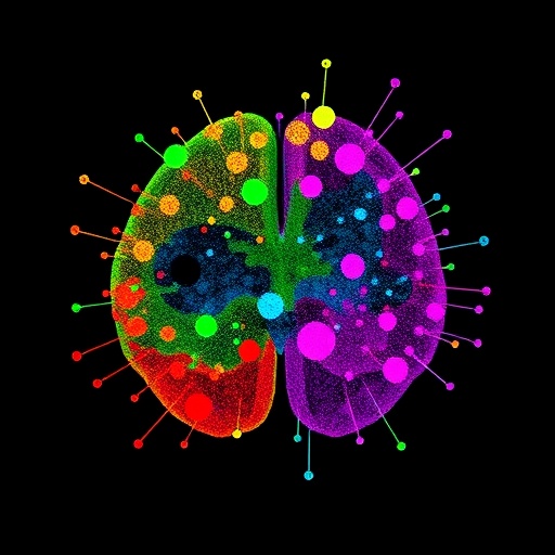
In a groundbreaking study that propels our understanding of head and neck squamous cell carcinoma (HNSCC) to new heights, researchers have employed cutting-edge single-cell RNA sequencing (scRNA-seq) technology to deeply profile the cellular landscape of lymph node metastases. This detailed molecular cartography reveals not only the complex heterogeneity of tumor cells but also unravels critical interactions within the immune milieu that accompany metastatic progression. The findings, recently published in Genes & Immunity, offer a compelling narrative on how tumor cells and their surrounding microenvironment evolve in the context of lymphatic spread, laying the groundwork for refined prognostic tools and novel therapeutic avenues.
HNSCC remains a formidable clinical challenge worldwide, marked by high rates of morbidity and mortality. Despite advances in therapeutic modalities, metastatic spread, particularly to lymph nodes, significantly worsens patient outcomes and complicates clinical management. Previous studies utilizing single-cell technologies have illuminated diverse components of the primary tumor microenvironment, including immune cell subpopulations and stromal elements. However, comprehensive dissection of metastatic lymph node tissues using similarly high-resolution techniques has lagged behind, leaving a critical gap in knowledge about metastatic niches in HNSCC.
Addressing this gap, the research team conducted an extensive comparative analysis involving scRNA-seq of seven metastatic lymph node samples alongside five non-metastatic lymph node controls derived from HNSCC patients. This rigorous design allowed researchers to pinpoint molecular and cellular alterations unique to the metastatic microenvironment. Their high-dimensional dataset unveiled alterations in both immune and stromal cells, highlighting cell types such as exhausted CD8+ T cells (CD8+ Tex), macrophages, and cancer-associated fibroblasts (CAFs) as key players reshaped during metastasis.
Strikingly, malignant cells exhibited pronounced heterogeneity, clustering into seven distinct transcriptional states. Among these, one cluster—cluster 2—emerged as a central hub of tumorigenic and metastatic gene expression programs. This cluster’s signature was enriched for pathways linked to cellular proliferation, invasion, and immune evasion, suggesting that it may serve as a driver population facilitating lymphatic dissemination. These insights underscore the complexity of tumor cells’ adaptive strategies in establishing metastatic colonies.
Immune cell subsets further underscored the intricate choreography within the metastatic node. Specific T cell subsets, including CD4 follicular helper cells expressing CXCL13 (CD4_Tfh_CXCL13) and regulatory T cells bearing ribosomal protein gene RPL26 (CD4_Treg_RPL26), were selectively expanded. Concurrently, cytotoxic CD8+ effector T cells marked by CREM, and natural killer cells high in GZMB expression (NK_GZMB) were also enriched in metastatic contexts, reflecting dynamic immune responses that may paradoxically both combat and facilitate tumor spread.
Macrophage populations flourished in metastatic nodes, with two distinct phenotypes — OLFML3-expressing and SPP1-expressing macrophages — being particularly prominent. These macrophage subsets possess immunomodulatory and extracellular matrix remodeling capabilities, likely aiding tumor cell invasion and sculpting an immune-suppressive niche that protects cancer cells from clearance. The presence of these specialized macrophage subsets reveals their nuanced contributions to the metastatic cascade.
A pivotal contribution of this study lies in the construction and validation of a novel prognostic model tailored for HNSCC patients, integrating molecular predictors derived from the single-cell data. Emblematic genes including INHBA, SFRP2, SPP1, and IFI27 serve as a composite biomarker panel. Elevated expression levels of these genes correlated robustly with disease aggressiveness and poorer overall survival, providing a refined metric for patient risk stratification that transcends conventional clinicopathologic parameters.
The prognostic model’s predictive accuracy was firmly tested across multiple patient cohorts, bolstering its utility in clinical settings. Its adoption could facilitate personalized therapeutic decision-making by identifying patients at heightened risk of metastasis who may benefit from intensified treatment regimens or closer surveillance. Additionally, the implicated genes themselves represent promising molecular targets for drug development aimed at curbing metastatic progression.
This study’s meticulous single-cell approach also illuminated the crosstalk between cancer-associated fibroblasts and immune cells within metastatic lymph nodes. CAFs, known architects of the tumor stroma, demonstrated gene expression changes modulating the extracellular matrix and secretion of immunoregulatory factors. Through these activities, CAFs likely orchestrate a permissive niche that fosters tumor growth and protects metastatic foci from immune attack.
On the technical front, the authors skillfully applied advanced bioinformatics pipelines to integrate and analyze the complex scRNA-seq data. Dimensionality reduction techniques and unsupervised clustering algorithms were critical in unraveling the distinct cellular states and inter-population relationships. The use of differential gene expression and pathway enrichment analyses elucidated the functional implications underpinning observed cellular phenotypes, enabling a comprehensive and mechanistic understanding of the metastatic microenvironment.
Beyond the immediate clinical relevance, this research exemplifies the transformative power of single-cell transcriptomics in cancer biology. By resolving tumor and immune heterogeneity at unprecedented resolution, it charts a path forward for the discovery of novel biomarkers and therapeutic targets across diverse malignancies. The approach adopted here could be seamlessly extended to other cancers prone to lymphatic metastasis, fostering a new era of precision oncology.
As this field evolves, integrating single-cell transcriptional maps with spatial transcriptomics and proteomics will further enhance our understanding of tumor ecosystems. Mapping the precise anatomical context of these cellular subsets and their interactions in situ will provide even richer insights into how metastatic niches evolve and respond to therapy. Such multimodal strategies hold the promise of revealing vulnerabilities that could be exploited to thwart metastatic progression more effectively.
In conclusion, the study by Wei, Zhang, Jiang et al. marks a significant milestone in oncological research, providing comprehensive insights into lymph node metastasis in HNSCC. Through an elegant combination of single-cell transcriptomics and sophisticated computational analysis, they have uncovered a cellular and molecular roadmap of metastasis that informs prognosis and offers new therapeutic targets. These findings embody the future of cancer research, where detailed cellular profiling transforms patient care and shapes next-generation treatments.
This pioneering work not only enriches our biological understanding but also holds practical implications for improving clinical outcomes in a notoriously challenging cancer type. It underscores the imperative of investigating metastatic microenvironments with single-cell precision and acts as a clarion call for continued investment in high-resolution multi-omics technologies. The promise of transforming HNSCC from a lethal disease to a manageable condition appears closer than ever.
As the scientific community applauds these advances, it is clear that unlocking the complexities of the tumor immune microenvironment at single-cell resolution will continue to drive innovation. The path illuminated by this research is fraught with complexity but also brimming with opportunity. With tools and insights such as these, the oncology field is poised to redefine metastasis research and ultimately improve the lives of patients worldwide.
Subject of Research: Lymph node metastasis and immune microenvironment heterogeneity in head and neck squamous cell carcinoma (HNSCC) analyzed via single-cell RNA sequencing.
Article Title: Single-cell transcriptomics reveals heterogeneity and immune microenvironment in lymphatic metastasis of head and neck squamous cell carcinoma.
Article References:
Wei, G., Zhang, G., Jiang, S. et al. Single-cell transcriptomics reveals heterogeneity and immune microenvironment in lymphatic metastasis of head and neck squamous cell carcinoma. Genes Immun (2026). https://doi.org/10.1038/s41435-026-00392-4
Image Credits: AI Generated
DOI: 16 March 2026
Tags: advanced molecular techniques in cancer researchheterogeneity of tumor cells in lymph nodesimmune cell diversity in cancer metastasisimmune microenvironment evolution in cancer spreadlymph node microenvironment in HNSCCmetastatic niche characterization in HNSCCmolecular profiling of metastatic cancer cellssingle-cell analysis of metastatic lymph nodessingle-cell RNA sequencing in head and neck cancersingle-cell transcriptomics for cancer prognosistherapeutic targets in head and neck squamous cell carcinomatumor-immune interactions in metastasis

