In a groundbreaking study that promises to reshape our understanding of cellular aging and joint health, researchers have presented a detailed comparative analysis of senescence processes within articular chondrocytes. These specialized cells, crucial for maintaining cartilage integrity, undergo aging through distinct mechanisms that have profound implications for degenerative diseases such as osteoarthritis. The investigation uniquely contrasts replicative senescence, a natural cellular aging process tied to DNA replication limits, with chemically-induced senescence, simulating accelerated aging under external stressors.
Articular chondrocytes are the primary cellular constituents of cartilage, the resilient and smooth elastic tissue cushioning the ends of bones in joints. As humans age, the functionality of these cells inevitably declines, contributing to cartilage degradation and joint diseases. Understanding the nuances that govern the senescence of chondrocytes is pivotal for developing targeted therapeutic interventions. This study dives deep into phenotypic changes—the observable characteristics—and molecular signatures that differentiate the two senescence pathways.
Replicative senescence occurs as cells progressively lose their proliferative capacity due to telomere shortening, a hallmark of cellular aging. On the other hand, chemically-induced senescence involves premature aging triggered by exposure to stressors such as reactive oxygen species, pro-inflammatory cytokines, or pharmacological agents. The researchers sought to elucidate whether these two modes of senescence share overlapping features or diverge distinctly, which could highlight different therapeutic targets in age-related cartilage diseases.
The research team deployed an array of advanced cytological assays combined with high-throughput molecular profiling techniques to track changes over time with unparalleled resolution. They meticulously documented alterations in cell morphology, senescence-associated β-galactosidase activity (a classical marker of senescence), and secretory profiles known as the senescence-associated secretory phenotype (SASP). This secretome, composed of inflammatory cytokines, matrix-degrading enzymes, and growth factors, plays a critical role in dictating the tissue environment and cellular crosstalk.
A major revelation emerged as the study delineated distinct molecular pathways activated in replicative versus chemically-induced senescence. While both conditions exhibited elevated SASP factors, the composition and intensity of secreted molecules diverged sharply. Replicative senescent chondrocytes predominantly showed an upregulation of anti-inflammatory mediators and tissue remodeling molecules, suggesting a more controlled senescence program. Conversely, chemically-induced senescence elicited a potent pro-inflammatory secretome, likely exacerbating cartilage matrix breakdown.
Epigenetic modifications, which govern gene expression without altering the DNA sequence, also displayed unique patterns between the two senescence types. The researchers utilized chromatin immunoprecipitation followed by sequencing to map histone modifications and DNA methylation landscapes. Replicative senescence involved gradual accumulation of repressive marks leading to transcriptional silencing of proliferation-related genes. Alternatively, chemically-induced senescence triggered abrupt epigenetic shifts that activated stress response pathways and inflammatory signaling.
Mitochondrial dynamics, central to cellular metabolism and reactive oxygen species generation, were differentially affected. Mitochondrial dysfunction is a well-documented driver of aging, and this study confirmed its prominent role in chemically-induced senescence, characterized by fragmented mitochondria and impaired oxidative phosphorylation. Interestingly, replicative senescent chondrocytes maintained relatively intact mitochondrial networks, hinting at alternative mechanisms underpinning their growth arrest.
The investigation further uncovered that DNA damage response (DDR) pathways were robustly engaged in both forms of senescence but with distinct kinetics and outcomes. Replicative senescence featured a slow and sustained activation of DDR markers, including γ-H2AX foci formation, indicative of persistent but manageable DNA lesions. In contrast, chemically-induced senescence manifested rapid and widespread DNA damage, overwhelming repair machineries and accelerating the onset of cellular dysfunction.
Importantly, this work also examined the role of cell cycle regulators such as p16^INK4a, p21^CIP1, and p53, which orchestrate the entry and maintenance of senescence. Despite common upregulation across both models, nuances in expression levels and regulatory feedback loops were evident, contributing to differences in senescent cell stability and potential reversibility. These findings hint at the possibility of fine-tuning senescence therapeutics by selectively targeting specific cell cycle pathways.
In an exciting translational angle, the study evaluated the responsiveness of senescent chondrocytes to senolytic drugs—compounds designed to selectively eliminate senescent cells. Chemically-induced senescent cells displayed greater sensitivity to these drugs, possibly due to their heightened pro-inflammatory state and metabolic vulnerabilities. In contrast, replicative senescent cells exhibited resistance, necessitating alternative strategies such as senostatic agents that suppress harmful SASP factors without cell elimination.
The research extends its relevance by highlighting how distinct senescence pathways influence cartilage homeostasis and disease progression differently in vivo. Animal models of osteoarthritis were employed to validate laboratory findings, demonstrating that chemically-induced senescence predominates under pathological conditions involving inflammation and oxidative stress, whereas replicative senescence contributes more to age-associated cartilage wear-and-tear. This duality underscores the complex interplay of internal and external aging stimuli on joint health.
Comprehensive RNA sequencing provided a wealth of transcriptomic data, enabling the identification of novel biomarkers and therapeutic targets. Genes involved in extracellular matrix synthesis, inflammation, and cell adhesion were differentially regulated, furnishing a molecular atlas of chondrocyte senescence states. Such data pave the way for precision medicine approaches to diagnose early senescence and intervene before irreversible joint damage occurs.
Given the societal burden of musculoskeletal disorders, this study’s implications resonate profoundly. By differentiating between replicative and chemically-induced senescence, it charts a nuanced path toward crafting personalized treatments that could alleviate pain and mobility loss in millions worldwide. Future research inspired by these findings might explore combinatory therapies that concurrently address both cellular aging mechanisms.
In essence, this investigation propels the field beyond a one-size-fits-all perception of cellular senescence, revealing a sophisticated landscape where the origin and nature of senescence dictate tissue destiny. By unlocking these molecular secrets, scientists are better equipped to engineer interventions that preserve cartilage integrity, delay osteoarthritis onset, and enhance quality of life for aging populations.
Overall, this study establishes a new paradigm in cellular aging research within joint tissues, blending phenotypic observations with deep molecular insights. Its comprehensive approach serves as a vital resource for the biomedical community, sparking renewed enthusiasm for senescence-targeted therapies and reinforcing the importance of experimental nuance in age-related disease modeling.
With these insights, the possibility emerges of extending the benefits of this research beyond cartilage, potentially influencing studies on other age-related disorders where senescence plays a critical role. The meticulous dissection of senescence pathways offers a powerful toolkit to decode cellular aging across tissues, heralding a future where age-associated degenerative diseases might be effectively managed or even prevented.
Subject of Research: Cellular senescence in articular chondrocytes and its implications for cartilage aging and osteoarthritis.
Article Title: Comparative phenotypic and molecular profiling of replicative and chemically-induced senescence in articular chondrocytes.
Article References:
Arteaga, M.B., Tarasova, K., Kidtiwong, A. et al. Comparative phenotypic and molecular profiling of replicative and chemically-induced senescence in articular chondrocytes.
Cell Death Discov. (2026). https://doi.org/10.1038/s41420-026-02961-y
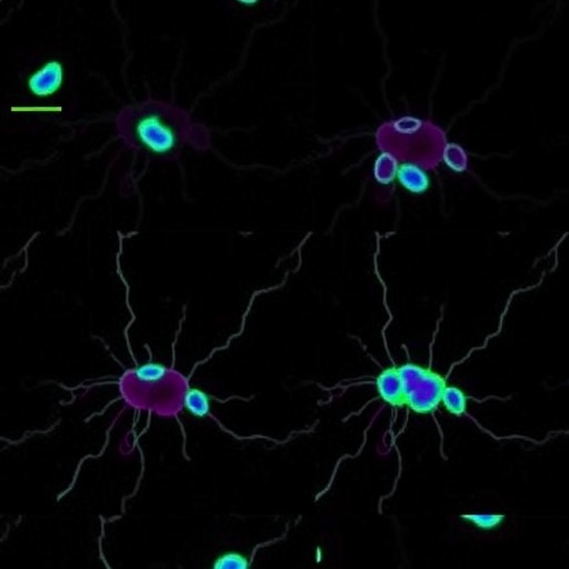
Image Credits: AI Generated
DOI: https://doi.org/10.1038/s41420-026-02961-y
Tags: articular chondrocyte senescence mechanismscartilage aging and degradationcellular aging pathways in joint healthcomparative analysis of senescence in cartilage cellsmolecular markers of chondrocyte agingosteoarthritis cellular pathologyoxidative stress-induced chondrocyte agingphenotypic changes in senescent chondrocytespro-inflammatory cytokines in cartilage degenerationreplicative versus chemically-induced senescencetargeted therapies for cartilage repairtelomere shortening in joint cells


