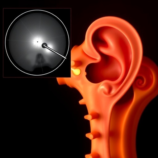
A groundbreaking advancement in medical imaging has emerged from the confluence of artificial intelligence and surgical innovation, promising to transform the landscape of cochlear implant surgeries. Researchers from St. Mary’s University, Trinity University, Vanderbilt University, and the Center for Advanced AI have unveiled a novel AI-driven technique capable of predicting the precise bone removal required in cochlear implant procedures using solely pre-operative computed tomography (CT) scans. This development addresses a long-standing challenge in otologic surgery: accurately anticipating the irregular and patient-specific shape of the mastoidectomy cavity created by removing part of the bone behind the ear.
Cochlear implants restore hearing by electrically stimulating the auditory nerve, and their successful implantation hinges on precise surgical access to the inner ear structures. Surgeons achieve this access via mastoidectomy, a procedure that entails the careful removal of mastoid bone. However, the absence of a consistent external boundary and the anatomical variations among individuals inject substantial complexity into preoperative planning and intraoperative navigation. Traditional imaging modalities and analysis tools have struggled to reliably delineate the intended mastoidectomy region, limiting surgeons’ ability to prepare accurately and increasing procedural risk.
The breakthrough described in the recent publication in the Journal of Medical Imaging represents a paradigm shift in how preoperative imaging data is leveraged to forecast surgical outcomes. The researchers overcame the inherent difficulty of manual annotation in post-surgical scans—known for their noise and variability—by deploying an advanced two-stage AI training strategy. The first stage harnesses the comparison between preoperative and postoperative CT data sets, enabling an unsupervised learning system to infer the extent and shape of bone removal. This stage exploits a mathematical approach focusing on structural similarities, allowing the AI to detect the transformed surgical anatomy without the need for finely detailed expert labels.
Building upon the preliminary predictions, the second AI model refines these approximations through the use of a three-dimensional loss function tailored by the Student-t distribution. This adaptation equips the algorithm to robustly contend with data imperfections, yielding enhanced accuracy and trustworthiness in the final output. This methodology illustrates the power of self-supervised learning frameworks in biomedical applications where pristine training data is scarce or infeasible to acquire, thereby opening new avenues for automating the interpretation of complex and irregular anatomical changes.
The robustness of this AI system was rigorously evaluated on a comprehensive dataset comprising 751 pairs of pre- and post-surgery CT scans. Benchmarking against a subset of 32 surgeon-annotated scans, the AI achieved a mean Dice coefficient of 0.72, surpassing the performance of several leading medical imaging models. The Dice score quantifies the congruence between predicted and actual bone removal shapes, with a higher score reflecting superior predictive fidelity. This metric validation underscores the system’s capability to precisely reconstruct the variable geometry surgeons encounter during mastoidectomy, offering a reliable computational surrogate for manual assessment.
Beyond prediction, the research team demonstrated the generation of full three-dimensional models that vividly represent the post-mastoidectomy bone surface, synthesized solely from the preoperative imaging input. Such 3D reconstructions hold immense potential in enhancing intraoperative visualization and guidance. Surgeons could rely on these models to navigate complex anatomical landscapes with heightened confidence, potentially integrating them with robotic surgical platforms or augmented reality interfaces. Additionally, these detailed digital replicas may serve an educational function, enabling trainees to gain a more nuanced understanding of operative anatomy and variations.
The implications of this work extend well beyond cochlear implant surgeries. The demonstrated AI training framework offers a blueprint for tackling similar challenges across numerous surgical domains where tissue alterations produce complex shapes hard to label manually. This adaptability could accelerate the development of predictive tools tailored to diverse interventions, smoothing the path toward personalized, precise, and safer surgical care. Furthermore, it addresses a critical bottleneck in medical imaging AI: reliance on meticulously curated annotations, which are often prohibitively time-consuming and inconsistently available.
Crucially, patient safety and surgical precision stand to benefit markedly from this innovation. By providing surgeons with accurate predictive insight into bone removal shapes before the operation, this technology can reduce unforeseen complications and streamline procedures. Such foresight is particularly valuable in settings lacking extensive expert resources, where access to advanced surgical planning tools can significantly enhance outcomes. As robotic-assisted and computer-navigated surgeries become more widespread, AI models like these will become indispensable components of the operating room ecosystem.
Despite its promising outcomes, the research team prudently acknowledges the necessity for broader clinical validation. Testing across multiple hospitals and diverse patient populations will be critical to ascertain the AI’s generalizability and robustness in varied clinical environments. The researchers are also exploring enhancements to the 3D reconstructions, aiming to imbue them with realistic textures and features that can improve surgeon interpretation and interaction during live procedures. These refinements are expected to fortify the tool’s utility and facilitate its adoption into routine clinical workflows.
This work opens exciting research frontiers in the intersection of AI and otologic surgery, suggesting a future where preoperative planning is augmented with precise, patient-specific anatomical predictions. By circumventing the limitations imposed by noisy or incomplete surgical imaging data, the approach showcases how machine learning can extract actionable intelligence from imperfect datasets. Such progress will likely inspire further innovations targeting other post-surgical morphological challenges, empowering clinicians with unprecedented foresight and control.
In summary, the fusion of sophisticated machine-learning algorithms with medical imaging has yielded a transformative approach to predicting mastoidectomy cavity shapes in cochlear implant surgery. This advanced method promises to enhance surgical safety, efficiency, and education by delivering accurate, label-free predictions solely from preoperative CT scans. As development continues and clinical trials expand, this AI-powered tool could become an indispensable asset in otologic surgery and beyond, heralding a new era of precision medicine facilitated by intelligent imaging technologies.
Subject of Research: Not applicable
Article Title: From preoperative computed tomography to postmastoidectomy mesh construction: mastoidectomy shape prediction for cochlear implant surgery
News Publication Date: 27-Jan-2026
Web References:
https://www.spiedigitallibrary.org/journals/journal-of-medical-imaging/volume-13/issue-1/014004/From-preoperative-computed-tomography-to-postmastoidectomy-mesh-construction–mastoidectomy/10.1117/1.JMI.13.1.014004.full
http://dx.doi.org/10.1117/1.JMI.13.1.014004
References: Y. Zhang et al., “From preoperative computed tomography to postmastoidectomy mesh construction: mastoidectomy shape prediction for cochlear implant surgery,” J. Med. Imaging 13(1), 014004 (2026), doi:10.1117/1.JMI.13.1.014004
Image Credits: Y. Zhang et al.
Keywords
Medical imaging; Inner ear; Cochlear implant surgery; Mastoidectomy; Artificial intelligence; Machine learning; Preoperative CT; 3D reconstruction; Post-surgery prediction; Self-supervised learning; Surgical planning; Anatomical modeling
Tags: advanced imaging techniques in cochlear implantationAI applications in otolaryngologyAI in medical imaging for ear surgeryAI-driven cochlear implant surgery planningAI-enhanced surgical navigation for cochlear implantsartificial intelligence in preoperative surgical planningbone removal accuracy in cochlear implant proceduresimproving cochlear implant surgical outcomes with AInovel AI techniques for mastoidpatient-specific mastoidectomy cavity predictionprecision bone removal in otologic surgerypreoperative CT scan analysis for mastoidectomy


