A Breakthrough in Brain Research: Soft 3D Electronic Meshes Unlock Neural Organoids’ Secrets
In a landmark advance poised to redefine neuroscience and regenerative medicine, researchers from Northwestern University, alongside scientists at the Shirley Ryan AbilityLab, have unveiled an innovative technology that captures the elusive electrical symphony within lab-grown human neural organoids. These organoid models—colloquially known as “mini brains”—are millimeter-sized, three-dimensional clusters derived from stem cells that replicate many aspects of human brain development and function. Until now, understanding their complex neural conversations has been limited by the constraints of existing measurement tools that could only probe a fraction of their neuronal network, leaving much of the brain-like activity a mystery.
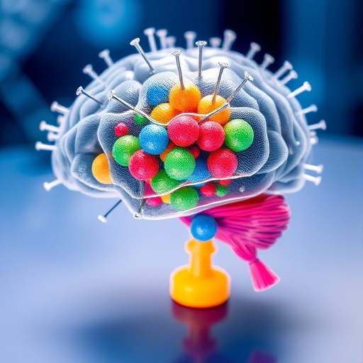
This frontier-shifting technology introduces a soft, highly porous 3D electronic mesh engineered to wrap seamlessly around the surface of neural organoids. Unlike conventional planar electrodes, which inadequately sample scattered regions, this mesh conforms precisely to the organoid’s contours, providing nearly complete spatial coverage with hundreds of micro-scale electrodes. Each electrode, as small as a single neuron—roughly 10 microns in diameter—acts as a portal into the dynamic electrical landscape of these developing neural circuits. This dense and responsive scaffold not only records but can also stimulate neuronal activity, bridging the gap from fragmentary glimpses to comprehensive, network-wide electrophysiological mapping.
The challenge addressed by this breakthrough lies in the fundamental geometry of organoids. Neural organoids develop into roughly spherical shapes with curved, intricate surfaces, embodying complex folded architectures reminiscent of actual brain tissue. Traditional microelectrode arrays, optimized for flat cell cultures, face a severe mismatch when applied to these three-dimensional shapes, leading to incomplete data and a loss of vital information. The Northwestern team solved this by designing a clever “pop-up book” style transformation: starting from a two-dimensional elastic lattice, they use mechanical buckling to convert the framework into a precisely engineered 3D scaffold. This transformation grants the mesh the ability to cradle the organoid gently, respecting its surface topography while maintaining porosity to support metabolic exchange critical for tissue health.
Crucially, the mesh’s design prioritizes tissue viability. Its highly perforated architecture allows oxygen and nutrients to permeate unhindered, ensuring organoids continue “breathing” throughout experimentation. This thoughtful integration of bioengineering principles was essential to avoid constraining the delicate tissues and to preserve authentic neural behavior during both recording and stimulation phases.
Upon enveloping the organoid, the mesh facilitates a remarkable leap in electrophysiological data acquisition. In configurations ranging up to an astounding 240 individually addressable electrodes covering 91% of the organoid’s surface, researchers detected rhythmic oscillations and network-wide electrical patterns previously inaccessible. The system’s fine spatial resolution coupled with temporal precision allowed scientists to visualize the propagation of activity waves across the entire 3D structure, uncovering intricate communication pathways within the developing neural ensemble.
Beyond passive observation, the team demonstrated the platform’s capability to modulate neural circuits actively. By delivering targeted electrical pulses through specific electrodes, they could evoke responses in designated regions, offering unprecedented control over the organoid’s electrical dynamics. When combined with advanced imaging techniques and optogenetic tools, this multi-modal approach sets a new benchmark for interrogating and manipulating miniature human neural networks in vitro.
The technology’s promise extends to pharmaceutical research and neurological disease modeling. Experiments testing various neuroactive compounds revealed distinct alterations in the neural network’s firing patterns. For instance, administration of 4-aminopyridine, a drug used clinically to improve motor function in multiple sclerosis, augmented neural signaling within the organoid. Conversely, exposure to botulinum toxin suppressed coordinated activity, illustrating the platform’s sensitivity in detecting pharmacological effects. These insights portend the utility of electrophysiologically interfaced organoids as living, human-relevant testbeds to accelerate drug discovery, safety assessment, and therapeutic development.
In an intriguing demonstration of bioengineering potential, researchers also showed that by modifying the mesh’s lattice design, they could influence organoid morphology. Non-spherical frameworks, including hexagonal and cubic shapes, guided neural tissues to grow into corresponding geometries. This controllable shaping capability hints at the future assembly of modular organoids, akin to biological “Lego blocks,” which could be combined to build complex, multi-organ systems for integrated physiology studies.
This advance is situated within a broader context of biomedical engineering and neuroscience, addressing a critical technological gap in organoid research at a time when these models are becoming central to disease modeling, personalized medicine, and regenerative strategies. John A. Rogers, a distinguished pioneer in bioelectronics, emphasizes that while stem cell-derived organoids have emerged as revolutionary models, their full potential remains stymied without hardware capable of comprehensive electrophysiological interrogation. The new mesh technology answers this call by enabling high-resolution, whole-network mapping and precise manipulation, thus unlocking new experimental horizons.
From a translational perspective, these advances open avenues for studying how complex brain disorders arise, testing drug efficacies in patient-specific contexts, and evaluating the restoration of coordinated neural function post-injury or disease. The ability to monitor and influence nearly all neurons in a mini brain model provides an unprecedented platform to deepen understanding of human brain physiology and pathology.
This pioneering work is supported by several funding bodies, including the National Institutes of Health, the National Science Foundation, and private foundations dedicated to neurorehabilitation and regenerative medicine. The study detailing this breakthrough, “Shape-conformal porous frameworks for full coverage of neural organoids and high-resolution electrophysiology,” was published in Nature Biomedical Engineering on February 18, 2026.
Looking forward, the research community is optimistic that combining innovations like this 3D electronic mesh with advancements in stem cell biology, imaging, and computational analysis will accelerate the trajectory toward personalized regenerative therapies. As organoid technology matures, equipped with tools for comprehensive interrogation and control, it promises to transform the biological and medical landscapes, bringing patient-specific neural tissue studies from the realm of possibility to practical reality.
Subject of Research: Human tissue samples
Article Title: Shape-conformal porous frameworks for full coverage of neural organoids and high-resolution electrophysiology
News Publication Date: 18-February-2026
Web References: https://www.nature.com/articles/s41551-026-01620-y
References: Rogers, J.A., Franz, C., Zhang, Y., Finan, J., et al. (2026). Shape-conformal porous frameworks for full coverage of neural organoids and high-resolution electrophysiology. Nature Biomedical Engineering. DOI: 10.1038/s41551-026-01620-y
Image Credits: John A. Rogers / Northwestern University
Keywords: Organoids, Brain tissue, Brain structure, Brain, Neural pathways, Nerve tissue, Nervous system, Bioelectronics, Biotechnology, Developmental biology, Neuroscience, Developmental disorders
Tags: 3D electronic mesh for neural organoidsbrain development research toolshuman mini brain modelsmicro-scale neuron electrodesmillimeter-sized brain organoidsneural circuit stimulation technologyneural organoid electrical recordingnext-generation bioelectronics interfaceNorthwestern University brain researchregenerative medicine neurotechShirley Ryan AbilityLab neurosciencesoft porous brain probes


