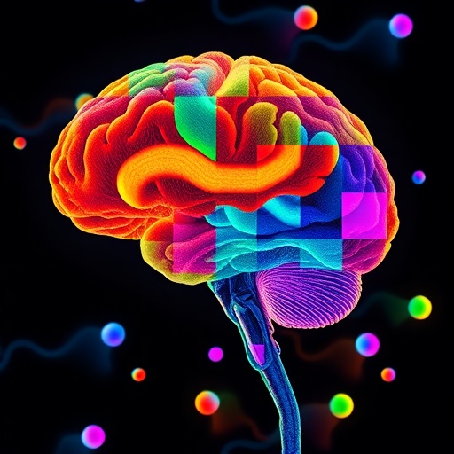
Understanding the brain’s complexity requires more than just knowing which genes are present; it demands insight into how proteins—the workhorses of the cell—are synthesized in distinct cellular environments. This challenge has long hampered neuroscience, as directly quantifying protein production, or translation, at the single-cell level in brain tissue remained elusive. Researchers from the University of California School of Medicine, Scripps Research, and their collaborators have unveiled a powerful new methodology named Ribo-STAMP, enabling precise mapping of protein synthesis across nearly 20,000 individual cells in the mouse hippocampus, a brain region pivotal to learning and memory. Their groundbreaking study was published in the prestigious journal Nature on February 18, 2026.
Gene expression is traditionally assessed by measuring levels of messenger RNA (mRNA), the intermediate molecule transcribed from DNA that encodes protein sequences. Although mRNA abundance has provided invaluable insights, especially with the rise of single-cell transcriptomics, it falls short in the brain. Neurons often sequester mRNAs within their extended processes, producing transcripts far in advance but strategically delaying their translation until needed. This spatial and temporal uncoupling generates a poor correlation between mRNA levels and actual protein synthesis, underscoring the necessity for direct measures of translation.
To address this gap, the team led by Dr. Gene Yeo developed Ribo-STAMP, an innovative approach that tags ribosomes—the molecular machines orchestrating translation—with a bespoke RNA-editing enzyme. As these ribosomes navigate mRNA strands, actively synthesizing proteins, the enzyme modifies the RNA in a distinctive manner. By analyzing these unique RNA edits through standard sequencing technologies, scientists can pinpoint exact mRNA molecules undergoing translation within single cells, overcoming previous technical limitations.
The application of Ribo-STAMP in the hippocampus revealed unanticipated translational profiles within cellular populations. Among neurons, two closely related subtypes known as CA1 and CA3 pyramidal cells exhibited strikingly different translation rates. CA3 pyramidal neurons displayed significantly elevated levels of protein synthesis compared to CA1. Given that both cell types are integral to memory processing circuits, this discovery suggests that differential translation could be a hitherto unrecognized layer of regulation governing how neuronal networks function and encode information.
Further insights emerged regarding mRNA isoforms—variant transcripts generated from the same gene differing in their untranslated regulatory regions. The study found that isoforms harboring longer regulatory domains correlated with increased translational efficiency in hippocampal neurons. This finding provides a molecular mechanism whereby alternative splicing or transcript variant selection may modulate protein output, influencing neuronal physiology and potentially contributing to pathological states when misregulated.
Beyond cell-type-specific differences, individual neurons exhibited dynamic “translation states,” fluctuating between high and low protein production activity. Neurons in a high translation state preferentially synthesized proteins associated with synaptic communication and energy metabolism, indicating that translation rates might serve as biomarkers of neuronal activity and engagement. Such states may reflect the functional heterogeneity and plasticity underlying complex brain processes.
The introduction of Ribo-STAMP represents a paradigm shift, enabling researchers to chart the “translatome” at single-cell resolution in complex tissues, bridging the crucial gap between transcriptomics and proteomics. This technology opens avenues to investigate how alterations in protein synthesis contribute to neurological disorders such as autism spectrum disorder, fragile X syndrome, and tuberous sclerosis complex—conditions linked to synaptic dysfunction and translational dysregulation.
Dr. Yeo underscores the transformative potential of this technique, emphasizing that it allows scientists to move beyond inferred protein production estimates toward direct, cell-specific translation measurements. This capability promises to enhance understanding of brain development, synaptic plasticity, and disease etiology by capturing the nuances of translational control in situ.
The research integrated expertise spanning molecular biology, neuroscience, and bioinformatics. Co-corresponding author Dr. Giordano Lippi from Scripps Research highlights how these findings disrupt prior assumptions regarding neuronal similarities and underscore the importance of investigating translation dynamics to comprehend the molecular underpinnings of memory circuits.
Notably, the large-scale dataset generated reveals a complex landscape in which translation efficiency varies not only across cell types but also within seemingly homogenous populations, suggesting that protein synthesis is finely tuned at multiple regulatory layers. Such detailed maps serve as invaluable resources for the neuroscience community to explore protein synthesis patterns in health and disease.
In their collaborative effort, the authors also explore the relationship between isoform selection and translation output, providing mechanistic insights with implications for understanding how transcript diversity impacts protein levels. This relationship could be crucial in unraveling the molecular basis of neurological disorders associated with aberrant splicing or transcript regulation.
With funding from the National Institutes of Health and contributions from numerous institutions, including the Broad Institute, Sanford Stem Cell Institute, and Houston Methodist Research Institute, this study exemplifies the power of interdisciplinary collaboration. The Ribo-STAMP methodology is poised to become a foundational tool in neuroscience research, illuminating the translational landscapes that define neuronal identity and function.
Looking ahead, researchers anticipate that applying Ribo-STAMP across various brain regions and disease models will shed light on translational dysregulation in neurodegeneration, developmental disorders, and synaptopathies. This technology not only deepens our molecular understanding of the brain but also holds promise for identifying novel therapeutic targets by pinpointing dysregulated protein synthesis at unprecedented detail.
Subject of Research: Protein synthesis (translation) mapping at single-cell resolution in the mouse hippocampus using Ribo-STAMP technology
Article Title: Scientists develop Ribo-STAMP to reveal single-cell protein production in the brain
News Publication Date: February 18, 2026
Web References:
Nature Study
Original Ribo-STAMP Method
References:
Yeo, G., Lippi, G., Sison, S., Kofman, E., Zampa, F., et al. (2026). Mapping translation in single cells reveals novel insights into neuronal protein synthesis in the hippocampus. Nature. https://doi.org/10.1038/s41586-026-10118-1
Image Credits: Erik Jepsen / UC San Diego
Keywords: RNA, Molecular genetics, Neurological disorders, Protein translation, Single-cell transcriptomics, Hippocampus, Neuronal protein synthesis
Tags: advances in neuroproteomics techniquesbrain diseases and protein translationdirect measurement of neuronal protein translationhippocampus protein production studymouse hippocampus single-cell analysisneuron mRNA translation regulationprotein synthesis in learning and memoryprotein synthesis mapping in brain cellsRibo-STAMP methodology neurosciencesingle-cell protein translation analysissingle-cell transcriptomics limitationsspatial-temporal control of protein synthesis

