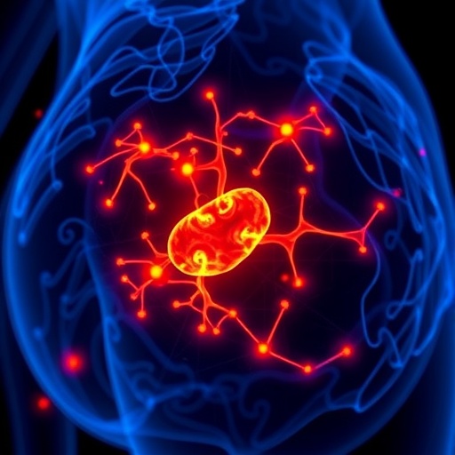
In a groundbreaking advancement poised to transform cancer research and diagnostics, a team of scientists led by A.J. Deloria and colleagues has introduced an innovative imaging technology dubbed Optical Coherence Photoacoustic Microscopy (OCPAM). This cutting-edge technique leverages the synergistic strengths of optical coherence tomography and photoacoustic microscopy to enable detailed three-dimensional imaging of cancer organoids. Their work, published in Light: Science & Applications, represents a significant leap forward in the ability to visualize complex tumor environments with unprecedented resolution and depth, providing a powerful new tool for oncological studies and personalized medicine.
The novelty of OCPAM lies in its dual-modality design, which integrates the high spatial resolution of optical coherence tomography (OCT) with the molecular specificity and penetration depth afforded by photoacoustic microscopy (PAM). OCT alone, while exceptional for capturing microstructural details, is often limited by poor chemical contrast and penetration depth in dense tissue. PAM complements this by employing pulsed laser excitation to generate ultrasonic waves via the photoacoustic effect, thus providing functional and molecular information that is otherwise invisible to traditional optical imaging. This fusion enables comprehensive visualization of cellular morphology co-registered with biochemical signatures, all in 3D.
Central to the significance of this technique is its application to three-dimensional cancer models developed from patient-derived organoids. These organoids are miniature, self-organized tumor-like tissue cultures that recapitulate key aspects of in vivo tumor microenvironments and heterogeneity. Historically, studying these 3D models has been hampered by the lack of imaging technologies that can delve deep into the tissue while maintaining high resolution and contrast. OCPAM bridges this gap, revealing intricate vascular structures, cellular arrangements, and biochemical landscapes crucial for understanding tumor biology and treatment response.
The researchers further enhance the utility of OCPAM through the integration of artificial intelligence-assisted analysis specifically tailored for organoid imaging. This AI framework automates the segmentation, classification, and quantification of various cellular and structural features within the complex 3D datasets. By combining advanced machine learning algorithms with rich multimodal imaging data, the system provides rapid, objective, and reproducible assessments, significantly accelerating data processing times and reducing observer bias—challenges that have long plagued manual histological examination.
From a technical standpoint, the OCPAM system employs a dual-laser configuration synchronized with an ultra-high sensitivity ultrasonic transducer array. The optical coherence module operates in the near-infrared spectrum, facilitating millimeter-scale imaging depths with micrometer lateral resolution. The photoacoustic component exploits a tunable laser source to excite chromophores such as hemoglobin and melanin, enabling real-time visualization of vascularization and pigmentation changes within the tumor microenvironment. Furthermore, the system’s scanning mechanism harnesses precision galvo mirrors, allowing rapid volumetric data acquisition at video-rate speeds without compromising image quality.
The integration of these modalities involves careful calibration to ensure co-registration of OCT and PAM images, aligning anatomical features with functional signals seamlessly. Post-acquisition, the volumetric datasets undergo advanced image reconstruction algorithms to enhance contrast and minimize artifacts, thereby maximizing diagnostic value. The accompanying AI pipeline was trained on extensive datasets comprising various organoid types and tumor compositions, enabling it to reliably discern cancerous from non-cancerous regions and extract quantitative metrics predictive of tumor aggressiveness and treatment sensitivity.
One of the striking advantages of OCPAM demonstrated in the study is its capability to monitor dynamic tumor behaviors in vitro over extended periods. Researchers showed that this platform could non-invasively track morphological changes, angiogenesis, and hypoxia-driven molecular shifts within organoids subjected to different chemotherapeutic agents. This opens new avenues for personalized medicine by facilitating real-time evaluation of drug efficacy at a cellular level, potentially guiding tailored therapeutic strategies that minimize toxicity while maximizing response.
In addition to cancer research, the versatility of OCPAM suggests broad applicability across biomedical fields requiring detailed 3D imaging of complex tissues. For example, its ability to visualize vascular networks and cellular heterogeneity could prove invaluable for studying developmental biology, tissue engineering, and neurodegenerative diseases where conventional imaging falls short. The system’s non-destructive nature further permits longitudinal studies on the same sample, preserving precious biological resources and enabling temporal investigations previously impractical.
The implications for clinical practice are equally promising. While current histopathological diagnosis relies heavily on invasive biopsies and time-consuming slide-based analysis, OCPAM could ultimately provide in situ, label-free imaging of biopsy specimens or surgical margins, aiding surgeons and pathologists with rapid, high-resolution feedback. Combined with AI-driven interpretation, this technology could reduce diagnostic errors, improve accuracy, and accelerate decision-making in oncologic care. The prospect of translating such a platform to bedside or intraoperative use could revolutionize cancer management paradigms.
The collaborative effort behind OCPAM involved multidisciplinary expertise spanning optical engineering, biomedical imaging, computational science, and oncology. This convergence underscores the growing recognition of cross-disciplinary approaches as key drivers of innovation. By uniting these domains, the team crafted a suite of solutions addressing longstanding challenges in tumor imaging—from fundamental physical constraints to practical hurdles in data analysis. Their success exemplifies the kind of integrative research that will undoubtedly shape the future landscape of medical technology.
Challenges remain, however, before OCPAM can be fully integrated into routine laboratory or clinical workflows. The complexity and cost of the dual-modality hardware may limit initial accessibility, necessitating further optimization and cost-reduction efforts. Moreover, comprehensive validation studies across diverse tumor types, including in vivo models and human clinical samples, are critical to establish robustness and generalizability. The AI algorithms also require ongoing refinement to adapt to novel imaging contexts and improvement in interpretability for clinical adoption.
Looking forward, the authors envision expanding the spectral capabilities of OCPAM to target multiple endogenous and exogenous contrast agents, further enriching molecular imaging capabilities. Coupling this with real-time AI decision support platforms could enable truly autonomous analysis pipelines capable of identifying subtle pathological changes earlier than currently possible. Integration with other imaging modalities such as fluorescence or Raman spectroscopy may also enhance diagnostic confidence by providing complementary biochemical insights.
In conclusion, the advent of Optical Coherence Photoacoustic Microscopy combined with AI-assisted organoid analysis marks a monumental step forward in cancer imaging technology. This hybrid approach addresses many of the limitations inherent to existing methods, offering detailed, multi-scale visualization and functional assessment of tumor structures in physiologically relevant 3D models. The ability to non-invasively probe tumor microenvironments with high resolution and rapid processing heralds new opportunities for understanding cancer progression, screening therapeutic responses, and eventually improving patient outcomes. As research and development continue, OCPAM holds immense potential to become a cornerstone platform in the era of precision oncology and beyond.
Subject of Research: The development and application of Optical Coherence Photoacoustic Microscopy (OCPAM) for high-resolution 3D imaging of cancer organoids combined with AI-assisted analysis to enhance understanding and evaluation of tumor biology and drug response.
Article Title: Optical coherence photoacoustic microscopy for 3D cancer model imaging with AI-assisted organoid analysis
Article References:
Deloria, A.J., Csiszar, A., Deng, S. et al. Optical coherence photoacoustic microscopy for 3D cancer model imaging with AI-assisted organoid analysis. Light Sci Appl 15, 106 (2026). https://doi.org/10.1038/s41377-025-02177-2
Image Credits: AI Generated
DOI: 05 February 2026
Tags: 3D Imaging of Cancer Organoidsadvanced cancer research technologiesAI-Enhanced Optical Coherence Photoacoustic MicroscopyDual-Modality Imaging TechniquesFunctional Imaging of CancerHigh Resolution Tumor VisualizationInnovative Imaging for Cancer TreatmentMolecular Imaging in Cancer ResearchOncological Studies with AIOptical Coherence Tomography in OncologyPersonalized Medicine and Cancer DiagnosticsPhotoacoustic Microscopy for Tumor Analysis

