In a groundbreaking fusion of advanced technologies and anthropological inquiry, researchers have unveiled new insights into the complexities of human pelvic anatomy, specifically focusing on the adult iliac auricular surface. This recent study, published in the International Journal of Legal Medicine, employs an innovative combination of geometric morphometrics and machine learning to dissect and interpret the nuanced asymmetries and sexual dimorphisms present in this critical anatomical structure. Such revelations promise to revolutionize forensic science and bioarchaeology by enhancing the precision with which human remains are analyzed and contextualized.
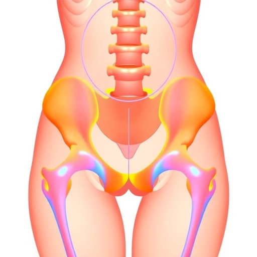
The iliac auricular surface, a key component of the pelvic girdle, functions mechanically as a critical interface between the ilium and sacrum, contributing to the load transfer between the spine and lower limbs. Variations in this region, including shape asymmetries and sexual dimorphisms, have long been recognized but remained challenging to quantify rigorously due to the subtlety of these features and the variability among individuals. Traditional morphometric approaches have typically relied on manual landmarking and subjective assessments, which can introduce bias and reduce reproducibility. However, the integration of computational geometric morphometrics marks a transformative advance.
Geometric morphometrics enables a detailed, quantitative description of shape by capturing landmark configurations and analyzing their spatial relationships. By applying this methodology to high-resolution 3D models of the iliac auricular surface, the research team could generate precise shape descriptors that represent individual variations in remarkable detail. This allowed them not only to discern consistent patterns of asymmetry but also to segregate these from sexual dimorphic traits with robust statistical power, an area that was previously fraught with interpretative challenges.
Machine learning algorithms further enhanced the analytical framework by automating pattern recognition and classification tasks that would be infeasibly complex for manual methods. The study employed supervised learning techniques to train models on labeled datasets, representing male and female specimens with known anatomical data. The algorithms demonstrated remarkable accuracy in distinguishing sexes based solely on the morphological parameters derived from the iliac auricular surfaces. This automated approach not only accelerates analysis but improves consistency, potentially creating a standardized protocol for forensic and anthropological applications worldwide.
The substantive findings reflect both biological reality and evolutionary history. Asymmetry detected in the auricular surfaces aligns with the concept of fluctuating asymmetry, a phenomenon often linked to developmental stability and environmental stresses during growth. The research elucidates how such asymmetries, though subtle, carry measurable significance and vary between sexes, offering deeper insights into pelvic biomechanics and reproductive biology. This may also have implications for understanding how pelvic shape influences locomotion and load-bearing capabilities, critical factors in both medical and evolutionary contexts.
Sexual dimorphism in the pelvis is well-documented, primarily reflecting the dual evolutionary pressures of childbirth and bipedal locomotion. This study’s sophisticated morphometric approach details the precise morphological differences in the iliac auricular surfaces, contributing to a more nuanced appreciation of how male and female pelvises differ not just overall but in localized shape characteristics. These differences, once quantified accurately, enhance sex estimation techniques vital for forensic identification, archaeological reconstructions, and even clinical assessments.
Importantly, the machine learning models were validated on diverse datasets, including specimens from multiple populations, addressing concerns of anthropological bias and ensuring broad applicability. This cross-population validation strengthens the utility of the findings and positions the method as a universally adaptable tool. By eradicating some of the subjective judgment inherent in traditional morphological classification, this research paves the way for more equitable and scientifically grounded approaches in bioarchaeology and forensic anthropology.
The combination of geometric morphometrics and machine learning also points toward new horizons in personalized medicine. Understanding the detailed morphology of the pelvis can inform clinical interventions, orthopedic surgery, and rehabilitation strategies tailored to individual anatomical variations. Furthermore, it can enhance biomechanical modeling used in prosthetics development, sports science, and injury prevention, showcasing the interdisciplinary potential of such technological integration.
On a theoretical front, these methods also open avenues for evolutionary biology to better track phenotypic changes across time and populations. By rigorously quantifying shape variations and asymmetries, scientists can investigate how environmental pressures, genetic factors, and lifestyle shifts impact pelvic morphology. This can lead to a richer narrative of human evolutionary adaptation, linking anatomical changes to functional and behavioral outcomes.
The study also underscores the potential of artificial intelligence to transform the way complex biological data is handled. By employing machine learning not as a mere analytical tool but as an integrated partner in discovery, the researchers highlight a shift in scientific methodology—one that emphasizes data-driven insights complemented by domain expertise. This model represents a paradigm shift, fostering enhanced reproducibility and setting a new benchmark for future morphological research.
While the research demonstrates impressive technological advancements, it also calls attention to remaining challenges. Precise 3D imaging acquisition, landmark selection criteria, and computational resource demands require careful optimization and standardization before routine implementation. Future work will need to address these aspects to create streamlined pipelines accessible to forensic practitioners and researchers alike.
Moreover, the societal implications of refining sex estimation techniques are profound. Ensuring ethical use of such technologies in legal and anthropological contexts necessitates careful consideration regarding privacy, consent, and cultural sensitivities. Transparent methodologies and open scientific dialogue will be critical to navigating these issues responsibly.
In conclusion, the study by Amendola, Navega, Barucci, and colleagues stands at the intersection of technology and anthropology, pushing the boundaries of what is possible in understanding human skeletal variation. By leveraging geometric morphometrics and machine learning to unravel the complex patterns of asymmetry and sexual dimorphism in the iliac auricular surface, this research offers a compelling vision of the future of forensic science, bioarchaeology, and evolutionary biology. The innovation and rigor presented promise not only to enhance scientific knowledge but also to impact practical applications that touch on identity, medicine, and history.
As these techniques become more refined and widespread, the potential for broader anthropological insights grows—offering a deeper understanding of human biology through the precise lens of shape, symmetry, and statistical subtlety. This novel approach heralds a leap forward in the science of human skeletal analysis, marrying computational power with anthropological acumen to decode the stories etched into our bones.
Subject of Research: Analysis of asymmetry and sexual dimorphism in the adult iliac auricular surface using geometric morphometrics and machine learning.
Article Title: Tracing asymmetry and sexual dimorphism in the adult iliac auricular surface: a geometric morphometrics and machine learning approach.
Article References:
Amendola, M., Navega, D., Barucci, A. et al. Tracing asymmetry and sexual dimorphism in the adult iliac auricular surface: a geometric morphometrics and machine learning approach. Int J Legal Med (2026). https://doi.org/10.1007/s00414-026-03726-z
Image Credits: AI Generated
DOI: https://doi.org/10.1007/s00414-026-03726-z
Tags: advancements in bioarchaeology methodsAI in pelvic anatomy researchanthropological insights from AIchallenges in traditional morphometrics.forensic applications of pelvic analysisgeometric morphometrics in anthropologyiliac auricular surface analysisinnovative technologies in anatomy studiesmachine learning in forensic scienceprecision in human remains analysisquantifying pelvic shape variationssexual dimorphisms in human anatomy


