In recent years, the complexity of vascular diseases has heightened the need for precise diagnostic tools to assess conditions such as carotid stenosis. This narrowing of the carotid arteries can lead to severe consequences, including strokes, thus compelling researchers to explore advanced imaging techniques that ensure both accuracy and efficacy. A pivotal study published in the journal “3D Print Med” delves into comparing the effectiveness of CT angiography against flow parameters using a hemodynamic phantom to evaluate carotid stenosis via duplex sonography. The research, led by a team of experts including Seidlová, Svoboda, and Voldřich, has opened up new avenues for understanding how different imaging modalities can help in the early detection and treatment planning for patients at risk of vascular complications.
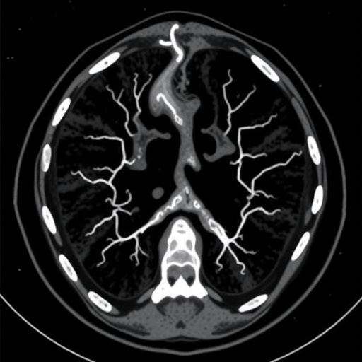
CT angiography has emerged as a preferred imaging modality because of its ability to provide detailed, high-resolution images of blood vessels. Unlike traditional angiography, which is more invasive and involves catheterization, CT angiography employs non-invasive techniques that can swiftly display the anatomy of the vascular structure. The capability to visualize the vascular system in three dimensions adds another layer of analysis that enhances the diagnostic process. However, its effectiveness in measuring flow parameters, especially in dynamic conditions within a phantom model of carotid stenosis, raises essential questions regarding its reliability compared to other standard techniques.
On the other hand, duplex sonography remains a cornerstone of vascular assessment due to its dynamic nature of real-time observation and continuous blood flow monitoring. This non-invasive method utilizes high-frequency sound waves to create images of blood vessels and assess blood flow. The integration of duplex sonography in the study provides a contrasting baseline to measure flow parameters effectively, which is essential for ruling out potential complications associated with carotid artery diseases. By combining these imaging techniques, researchers aim to provide a comprehensive perspective on the diagnostic capabilities available for assessing vascular conditions.
The study employed a sophisticated hemodynamic phantom model simulating carotid stenosis, allowing researchers to replicate various conditions reflecting real-life scenarios. This innovative model facilitated precise measurements of blood flow dynamics, which is critical for validating the performance of the imaging techniques being compared. Through this approach, the researchers aimed to establish correlations between the imaging data obtained from CT angiography and the hemodynamic parameters assessed by duplex sonography. The implications of such findings could enhance clinical decision-making, leading to better patient outcomes and improved management strategies for carotid artery diseases.
As the research delved deeper into the comparative analysis, it became evident that discrepancies exist between the information garnered from both imaging modalities. CT angiography might reveal structural abnormalities and the overall morphology of the arteries but could fall short when it comes to real-time blood flow evaluations. Conversely, duplex sonography excels in capturing hemodynamic variations but may sometimes lack the detailed anatomical precision offered by CT imaging. This duality highlights the necessity for a balanced approach in utilizing both techniques in clinical practice to achieve a comprehensive assessment of carotid stenosis.
In terms of clinical relevance, understanding the strengths and limitations of both imaging modalities can directly influence treatment protocols and patient management. Decision-makers in healthcare settings can benefit significantly from this type of comprehensive investigative work, as they are tasked with ensuring that the most appropriate diagnostic measures are employed to guide their clinical decisions. As a result, further exploration through well-structured studies becomes indispensable to enhance the existing knowledge base and refine the approaches towards diagnosing vascular diseases.
Moreover, the use of advanced hemodynamic phantoms provides a robust platform for future research initiatives. As the landscape of vascular imaging continues to evolve with technology, the ability to recreate human physiological conditions in lab settings offers unparalleled opportunities to investigate and refine diagnostic protocols. This advancement stands to benefit not just researchers but also practitioners in the field, ultimately contributing to better patient care and outcomes through more informed decisions.
As the findings from this comparative study make their way into the broader scientific community, one anticipates a ripple effect that encourages further research into multimodal imaging approaches. The quest for a more holistic strategy to evaluate vascular health may pave the way for integrated systems that harness the strengths of various imaging techniques while mitigating their weaknesses. Embracing such innovations could revolutionize how conditions like carotid stenosis are diagnosed and managed, ultimately leading to enhanced preventive strategies against strokes and other vascular maladies.
Moving forward, it will be crucial for multidisciplinary collaborations between imaging specialists, vascular surgeons, and researchers to synthesize knowledge from various specializations. This collaborative spirit will foster an environment conducive to pushing boundaries and improving patient care efficiency and effectiveness. Initiatives aimed at rounding out the understanding of vascular pathologies through comprehensive imaging analyses hold the potential to yield groundbreaking insights and ultimately revolutionize care modalities in cardiovascular medicine.
In conclusion, as the field of medical imaging matures, honing in on comparative studies, such as the one led by Seidlová and colleagues, becomes essential for contours defining advancements and innovations in vascular treatment protocols. The evolution of diagnostic techniques, driven by empirical research and technological growth, ensures that healthcare providers remain better equipped to address complex conditions like carotid stenosis. Such studies not only augment current clinical practices but also lay the groundwork for future explorations that promise to enhance both diagnostic accuracy and therapeutic outcomes.
In summation, the comparative exploration of CT angiography and duplex sonography through a hemodynamic phantom model has shed light on vital imaging fronts in the management of carotid stenosis. As advancements in technology unfold, the need for such thorough evaluations becomes ever more significant—aiming ultimately to improve the healthcare landscape concerning vascular health and disease management.
Subject of Research: Comparative study of imaging techniques for carotid stenosis evaluation.
Article Title: A comparative study between CT angiography and flow parameters in hemodynamic phantom of carotid stenosis evaluated by duplex sonography.
Article References:
Seidlová, A., Svoboda, N., Voldřich, R. et al. A comparative study between CT angiography and flow parameters in hemodynamic phantom of carotid stenosis evaluated by duplex sonography.
3D Print Med 11, 29 (2025). https://doi.org/10.1186/s41205-025-00271-0
Image Credits: AI Generated
DOI: https://doi.org/10.1186/s41205-025-00271-0
Keywords: Imaging, Carotid Stenosis, CT Angiography, Duplex Sonography, Hemodynamic Phantom, Vascular Diseases, Diagnostic Techniques, Comparative Analysis, Patient Care, Stroke Prevention, Blood Flow Dynamics, Medical Imaging, Collaborative Research, Vascular Health.
Tags: 3D imaging in vascular assessmentadvanced imaging techniques for strokescomparison of imaging modalities for stenosisCT angiography for carotid stenosisduplex sonography in vascular imagingearly detection of carotid artery narrowingeffectiveness of CT angiography vs duplexflow parameters in carotid stenosishemodynamic phantom in vascular researchnon-invasive carotid artery evaluationprecision diagnostics in vascular diseasesresearch on vascular complications detection.
