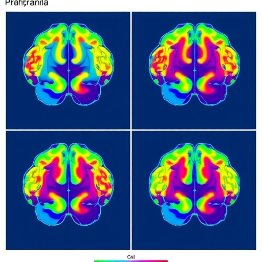
In a groundbreaking study published in Nature, researchers have unveiled how the dorsomedial prefrontal cortex (dmPFC) intricately encodes information about environmental cues to drive goal-directed behavior in mice. This neural mechanism sheds unprecedented light on the brain’s ability to distinguish and prioritize motivationally relevant stimuli, critical for balancing approach and avoidance behaviors essential to survival. The study innovatively disentangles three fundamental dimensions governing stimulus relevance—salience, valence, and value—revealing a sophisticated and dynamic population coding within dmPFC networks.
Our interaction with the environment hinges on extracting meaningful signals that signal potential threats or rewards. The dmPFC, a brain region pivotal in higher cognitive functions, occupies a central role in translating these sensory inputs into motivated actions. Despite its acknowledged importance, how this area processes complex cue information—specifically how it segregates and integrates salience (importance), valence (positive or negative emotional charge), and value (expected outcome magnitude)—has remained a mystery until now. By harnessing calcium imaging technology in freely behaving male mice, Winke, Lüthi, Herry, and colleagues achieved an unprecedented granularity in observing single-neuron activity as the animals learned to discriminate stimuli predicting varying outcomes of reward or punishment.
The methodology leveraged in this study represents a major advancement. Using calcium indicators that fluoresce upon neuronal activation, the researchers tracked population-level dmPFC neural dynamics longitudinally across associative learning phases. This approach permitted real-time, cellular-resolution mapping of how neurons encode stimulus features relevant to motivational salience and affective value. By presenting mice with cues linked to either positive (appetitive) or negative (aversive) outcomes, the study created a framework enabling the dissociation of neural representations for salience, valence, and value—dimensions often conflated in previous investigations.
One of the study’s central findings concerns the geometrical organization of neuronal activity within the dmPFC. Neuronal populations were found to encode appetitive and aversive values along distinct, orthogonal axes—essentially in separate neural “spaces.” This segregation allows simultaneous but independent processing of positive and negative motivational aspects of stimuli, preventing ambiguity in the organism’s behavioral response. Notably, subsets of neurons within the dmPFC were specialized to encode either valence or the stimulus’ salience, highlighting a multifaceted coding strategy that supports adaptive decision-making. Such orthogonality in neural representation represents a novel architectural principle for the neural basis of motivated behaviors.
Equally revealing was how these neural representations evolved dynamically during learning. Initial generalized responses to environmental cues sharpened over sessions, with specific dmPFC populations emerging to distinctly encode the learned value associated with each stimulus. This plasticity suggests that the dmPFC does not merely relay sensory information but actively configures its networks to optimize behavior by encoding motivational parameters in well-defined formats. This learning-related neural geometry ensures that the brain’s motivational circuits can flexibly adjust focus between reward-seeking and threat avoidance, based on contextual cue significance.
Beyond basic neuroscience, these findings have profound implications for conceptualizing the neural underpinnings of complex cognitive functions. Disorders such as anxiety, addiction, and depression often involve maladaptive valuations of environmental stimuli—where either threatening signals are exaggerated or rewarding cues are undervalued. Understanding how the dmPFC orchestrates the representation of motivational dimensions opens new avenues for targeted interventions that could recalibrate disordered neural coding patterns. The study’s insights into the geometry of neural representations provide a platform for developing biomimetic models that simulate decision-making disruptions observed in psychiatric illnesses.
From a broader evolutionary perspective, the demonstrated population coding mechanisms align with theories that prefrontal cortex networks evolved to handle complex, multidimensional valuation computations essential for survival. Encoding valence, salience, and value along distinct neural channels may confer computational advantages, allowing organisms to rapidly prioritize behaviors when confronted with competing stimuli in unpredictable environments. The exquisite tuning of dmPFC neurons to motivationally salient cues highlights the sophistication of mammalian cognition, bridging sensorimotor integration with affective processing.
Importantly, the researchers emphasize that the multifunctional nature of the dmPFC neural ensembles enables concurrent processing of opposing motivational forces rather than a simplistic linear summation. This nuanced balance allows animals to dynamically bias their behavior towards either approach or avoidance depending on the perceived cost-benefit scenario. Consequently, the study’s population-level perspective contrasts with earlier paradigms that focused mainly on single-neuron responses, providing a more accurate depiction of the emergent properties underpinning motivated behavior.
Technological advancements in calcium imaging played a critical role here, as traditional electrophysiological recordings could not feasibly resolve the high-dimensional activity patterns across broad dmPFC populations in freely moving subjects. The researchers’ innovative integration of behavioral paradigms with in vivo imaging permitted a granular yet ecologically valid examination of associative learning processes, devoid of confounds introduced by restraint or anesthesia. This methodological synergy sets a new benchmark for investigating neural computations underlying complex behaviors.
The research team also suggests that these findings may extend to understanding human prefrontal cortex functionality. Given the conserved architecture and evolutionary continuity of prefrontal regions across mammals, insights gained from murine models offer translational value. Dissecting how valence and value are represented geometrically in human prefrontal networks could improve cognitive-behavioral approaches that depend on modifying incentive processing, such as therapies for addiction or mood disorders.
In conclusion, this pioneering study elucidates how the dmPFC encodes learned environmental cues in a high-dimensional neural space structured by the orthogonal encoding of salience, valence, and value. Such dynamic neural geometry governs the delicate balance of motivated behavior, enabling organisms to efficiently navigate complex environments by judiciously weighing opportunities and threats. The findings redefine our understanding of prefrontal cortex functionality and establish a framework for future research aimed at unraveling the neural algorithms behind decision-making and emotional regulation.
As neuroscience continues to unravel the neural language of behavior, this research represents a milestone demonstrating the intricate cortical choreography that underlies even the simplest decisions. By capturing the population-level dynamics of neuronal ensembles, the authors illuminate principles that could inspire artificial intelligence models seeking to emulate adaptive, value-based behaviors. Ultimately, this work exemplifies how advanced neurotechnologies can deepen our grasp of brain function and its role in shaping motivated actions fundamental to life itself.
Subject of Research: Neural Encoding of Motivational Cue Dimensions in the Dorsomedial Prefrontal Cortex
Article Title: Prefrontal neural geometry of learned cues guides motivated behaviours
Article References:
Winke, N., Lüthi, A., Herry, C. et al. Prefrontal neural geometry of learned cues guides motivated behaviours. Nature (2026). https://doi.org/10.1038/s41586-025-09902-2
Image Credits: AI Generated
DOI: https://doi.org/10.1038/s41586-025-09902-2
Keywords: dorsomedial prefrontal cortex, dmPFC, motivated behaviour, salience, valence, value coding, calcium imaging, neural population dynamics, associative learning, neural geometry, reward, punishment, decision-making
Tags: advancements in neuroscience methodologiesapproach avoidance behavior mechanismscalcium imaging in neurosciencedmPFC and cognitive functionsenvironmental cues and motivationgoal-directed behavior in micemotivationally relevant stimuli processingmouse models in behavioral researchprefrontal cortex neural codingsalience valence and value in decision makingsingle-neuron activity observationstimulus relevance and survival

