In a groundbreaking advancement poised to redefine the landscape of fluorescence microscopy, a team of researchers led by Wang, Z., Gu, W., and Xu, S. has unveiled a novel computational approach to decouple the point spread function (PSF) from the imaging process. This pioneering work, recently published in Light: Science & Applications, offers a transformative pathway to enhance the resolution and accuracy of fluorescent imaging, a crucial tool in biological and medical sciences.
Fluorescence microscopy has long been celebrated for its ability to illuminate the intricate details of biological specimens by tagging molecules with fluorescent markers. However, the technology’s precision is inherently limited by the point spread function, an optical artifact that describes how a microscope blurs a point source of light. The PSF acts akin to a fingerprint of the microscope’s optics but often convolutes the true signal with noise and distortion, posing persistent challenges in image reconstruction and analysis.
The research team’s revolutionary strategy centers on computational decoupling of this function, effectively disentangling the blur induced by the optical system from the true fluorescence emission of the sample. By leveraging advanced algorithms and mathematical models, they have developed a robust framework that separates the PSF from measured images, thus refining the final output with remarkable clarity and detail.
At the core of this approach is a sophisticated algorithm that models the PSF as an independent variable rather than a static characteristic embedded in the optical setup. This shift in perspective allows for dynamic adjustment and correction of the PSF during image processing, adapting to variations inherent in biological samples and experimental conditions. Such adaptability marks a significant leap beyond conventional methods that often assume a fixed PSF, limiting the fidelity of the reconstructed images.
The implications of this computational feat are profound. Enhanced resolution in fluorescence microscopy translates directly into the ability to observe cellular structures and interactions with unprecedented precision. For instance, researchers can now more accurately map the location and behavior of proteins within cells, track dynamic processes like gene expression or signal transduction, and identify subtle pathological changes that underpin diseases.
Moreover, this technique does not necessitate modifying existing hardware but rather complements current fluorescence microscopes through software upgrades. This accessibility means widespread adoption could be realized swiftly across labs worldwide, democratizing high-resolution imaging and accelerating discoveries across various disciplines including neurobiology, oncology, and developmental biology.
The researchers demonstrated the efficacy of their method using a variety of biological samples, ranging from cultured cells to complex tissues. In each case, the decoupling algorithm significantly sharpened image quality, unveiled finer structural details, and improved the signal-to-noise ratio. These results were validated against established benchmarks, confirming the method’s accuracy and reliability.
Importantly, the team addressed computational efficiency, a crucial consideration for real-time or high-throughput microscopy applications. Their algorithm is optimized to run on widely available computing infrastructure while maintaining a balance between processing speed and reconstruction fidelity. This makes it feasible to integrate their solution into live imaging setups, where timely feedback is essential.
Beyond practical applications, this work opens new avenues for theoretical exploration in optics and computational imaging. By treating the PSF as a dynamically estimable entity, it challenges longstanding assumptions in optical physics and paves the way for further innovations that could extend into other imaging modalities such as super-resolution and multiphoton microscopy.
The publication also highlights the collaborative synergies between computational scientists, optical physicists, and biologists, underscoring how interdisciplinary approaches can tackle long-standing challenges in scientific instrumentation. This fusion of expertise culminated in an elegant yet powerful solution that promises to elevate the fidelity of biological imaging and deepen our understanding of the microscopic world.
As fluorescence microscopy continues to be instrumental in unraveling cellular mechanisms and biophysical processes, the ability to computationally decouple and refine the PSF may well become a standard feature of tomorrow’s imaging toolkits. This advancement not only enriches the quality of visual data but also enhances the interpretability and quantitative accuracy, essential for translational research and clinical applications.
Looking ahead, ongoing research aims to extend this framework to more complex imaging scenarios, including live-cell dynamics under varying environmental conditions. Efforts to integrate machine learning approaches for automated PSF estimation and correction are also underway, which could further amplify the algorithm’s robustness and usability.
This innovation, detailed meticulously in their paper, represents a milestone in microscopy technology by offering a computational resolution to optical limitations that have challenged scientists for decades. It exemplifies how computational techniques are increasingly becoming as vital as physical hardware improvements in advancing scientific frontiers.
The marriage of computation and optics in this work heralds an exciting era where conventional trade-offs between image resolution, speed, and practicality are being redefined. As researchers worldwide embrace and build upon these findings, fluorescence microscopy stands on the cusp of unprecedented precision and versatility, enabling new discoveries that were once beyond reach.
Wang, Z., Gu, W., Xu, S., and their colleagues have thus carved a path towards a future where imaging technology is more agile, responsive, and accurate. Their contribution extends the boundaries not just of microscopy, but of how we perceive and visualize the fundamental structures of life itself.
Subject of Research:
Optical and computational enhancement of fluorescence microscopy through point spread function decoupling.
Article Title:
Point spread function decoupling in computational fluorescence microscopy.
Article References:
Wang, Z., Gu, W., Xu, S. et al. Point spread function decoupling in computational fluorescence microscopy. Light Sci Appl 15, 26 (2026). https://doi.org/10.1038/s41377-025-02112-5
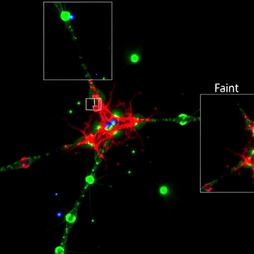
Image Credits: AI Generated
DOI: 10.1038/s41377-025-02112-5
Keywords:
Fluorescence Microscopy, Point Spread Function, Computational Imaging, Image Reconstruction, Optical Resolution, Biological Imaging, Fluorescence Signal Processing, Microscopy Algorithms, Computational Optics
Tags: biological specimen analysiscomputational imaging techniquesdecoupling point spread functionsfluorescence microscopy advancementsfluorescent marker technologyimage reconstruction methodsmathematical models in microscopynoise reduction in fluorescence imagingoptical artifacts in imagingresearch in biological imagingresolution enhancement in microscopytransformative microscopy approaches


