In the relentless quest to confront neurodegenerative diseases, a groundbreaking study has recently emerged, revealing novel insights into the early, often hidden, decline of brain health associated with isolated REM sleep behavior disorder (iRBD). This disorder, characterized by the loss of muscle atonia during REM sleep, frequently precedes synucleinopathies such as Parkinson’s disease and dementia with Lewy bodies, offering a critical window for early intervention. However, the clandestine nature of neurodegeneration in iRBD has long posed a formidable challenge to clinicians and researchers alike. Now, an innovative approach combining advanced MRI microstructural imaging with assessments of glymphatic flow has begun to peel back the veil, uncovering subtle neural alterations that have remained undetected until this moment.
Traditional neuroimaging modalities have struggled to capture the subtle microstructural changes occurring in the brains of iRBD patients. Yet, through the lens of state-of-the-art diffusion MRI techniques, researchers have succeeded in dissecting the intricate architecture of neural tissue with unprecedented precision. By mapping variations in fractional anisotropy and mean diffusivity across strategic brain regions, the team identified microstructural aberrations that signal the onset of neurodegenerative processes well before they manifest clinically. These changes offer vital clues about the neuronal pathways most vulnerable in the early stages of iRBD, highlighting the insidious progression occurring beneath the surface.
Complementing this microstructural exploration is the pioneering assessment of the brain’s glymphatic system—a vital clearance mechanism responsible for the removal of metabolic waste products from the central nervous system. Emerging evidence positions glymphatic dysfunction as a key player in the pathogenesis of neurodegenerative disorders. Leveraging advanced MRI sequences sensitive to cerebrospinal fluid dynamics, the researchers quantified glymphatic flow efficiency, unveiling significant impairments in patients with iRBD. This finding not only underlines the systemic nature of the disease but implicates glymphatic insufficiency as a potential driver of toxic protein accumulation, thus accelerating neurodegenerescence.
The study’s integrated methodology underscores the importance of a multimodal approach in unraveling the complexities of neurodegeneration in prodromal disease stages. By correlating microstructural disruptions with compromised glymphatic clearance, the research team has constructed a compelling narrative linking structural degradation to functional impairment within the brain’s clearance pathways. This convergence bolsters the hypothesis that early neurodegeneration in iRBD is a multifactorial process, weaving together tissue architecture breakdown and diminished neurotoxic waste removal.
Remarkably, these findings offer a beacon of hope for early diagnosis and therapeutic intervention. Detecting microstructural and glymphatic alterations prior to overt symptomatology could enable clinicians to stratify patients based on their neurodegenerative risk profile, facilitating personalized medicine approaches. Moreover, this paradigm may pave the way for innovative treatments aimed at enhancing glymphatic function, potentially decelerating or halting the progression of synucleinopathies before irreversible damage ensues.
In light of these revelations, the implications for clinical practice are profound. Routine incorporation of sophisticated MRI protocols targeting microstructural markers and glymphatic dynamics might become instrumental in the early identification of individuals poised to develop Parkinsonian syndromes. Such diagnostic precision aligns with the overarching goal of neuroprotective strategies: intercepting disease progression at the earliest possible juncture when interventions are most efficacious.
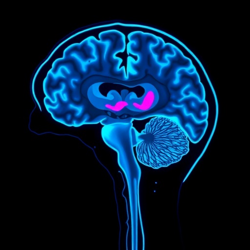
The research further elucidates the regional specificity of neurodegenerative alterations in iRBD, with particular vulnerability noted in subcortical nuclei and associated white matter tracts. These regions, integral to motor control and cognitive function, are critical nodes within neural networks susceptible to synucleinopathy-induced disruption. The concordance between microstructural compromise in these areas and diminished glymphatic clearance suggests a pathophysiological cascade that precipitates synaptic failure and neuronal loss.
From a technical perspective, the deployment of advanced diffusion MRI models, such as neurite orientation dispersion and density imaging (NODDI), alongside time-resolved glymphatic flow measurements, exemplifies the cutting-edge imaging arsenal propelling this field forward. These tools afford unparalleled resolution in capturing the subtle microenvironmental changes within the brain, facilitating a granular understanding of disease evolution. The fidelity of these imaging biomarkers sets a new standard for neurodegeneration research, potentially extending beyond iRBD to a broader spectrum of neurological disorders.
Moreover, the study addresses a pivotal gap in the understanding of how the glymphatic system’s impairment interplays with proteinopathy in neurodegenerative diseases. The authors hypothesize that deficient clearance mechanisms exacerbate the accumulation of alpha-synuclein aggregates, thereby perpetuating a vicious cycle of neuronal toxicity and inflammation. This hypothesis resonates with emerging models that integrate vascular, inflammatory, and proteostatic pathways to comprehensively explain neurodegeneration.
Notably, this research also underscores the dynamic interrelation between sleep physiology and glymphatic activity, anchoring the significance of REM sleep behavior in maintaining neural homeostasis. The disruption of muscle atonia characteristic of iRBD might reflect, or even contribute to, disturbed glymphatic pumping mechanisms dependent on cyclical cerebrospinal fluid fluxes during healthy sleep cycles. This bidirectional relationship opens intriguing possibilities for therapeutic modulation of sleep architecture as a means to bolster glymphatic clearance.
The validation of these findings across larger cohorts and longitudinal studies will be essential to ascertain the prognostic utility of MRI-derived microstructural and glymphatic markers. If corroborated, such biomarkers could revolutionize clinical trial design by providing objective surrogate endpoints that reflect disease biology rather than relying solely on clinical symptom progression. This shift promises to accelerate the pipeline for novel therapies targeting early neurodegeneration.
Furthermore, the translational potential of this research extends into the realm of neuroprotective drug development. Compounds aiming to enhance glymphatic system performance or protect microstructural integrity could be screened and monitored using these advanced imaging biomarkers, fostering a precision-medicine framework that tailors treatment to individual pathophysiological profiles.
The study’s meticulous design and rigorous analytical framework serve as a testament to the synergy between clinical neurology, neuroimaging physics, and neuropathology. It heralds a new era in which the veil obscuring early neurodegenerative changes in prodromal disorders like iRBD is lifted, unveiling actionable insights that bridge the gap between bench research and bedside application.
In sum, this landmark investigation not only deepens our understanding of the neurobiological underpinnings of isolated REM sleep behavior disorder but also charts a transformative course toward earlier detection and intervention in neurodegenerative diseases. Its integration of microstructural MRI and glymphatic flow analysis exemplifies the innovative spirit needed to confront the rising tide of neurodegenerative disorders with fresh eyes and potent tools.
As the global population ages and the burden of Parkinsonian disorders escalates, studies such as this will be crucial in shifting the paradigm from reactive treatment to proactive prevention. By capturing the silent whispers of neurodegeneration before they crescendo into clinical manifestations, medicine moves closer to subverting disease and preserving the evolving complexity of the human brain.
Subject of Research:
Neurodegeneration in isolated REM sleep behavior disorder through advanced MRI microstructural imaging and glymphatic system evaluation.
Article Title:
Unveiling hidden neurodegeneration in isolated REM sleep behavior disorder through MRI microstructure and glymphatic flow.
Article References:
Basaia, S., Sarasso, E., Gardoni, A. et al. Unveiling hidden neurodegeneration in isolated REM sleep behavior disorder through MRI microstructure and glymphatic flow. npj Parkinsons Dis. 11, 346 (2025). https://doi.org/10.1038/s41531-025-01193-8
Image Credits:
AI Generated
DOI:
https://doi.org/10.1038/s41531-025-01193-8
Tags: advanced MRI techniques for neuroimagingclinical challenges in diagnosing iRBDdiffusion MRI and neural tissue architectureearly intervention in neurodegenerative diseasesfractional anisotropy in iRBD patientsglymphatic flow and brain healthisolated REM sleep behavior disordermean diffusivity and neurodegenerative processesmicrostructural imaging in neurodegenerationneurodegeneration in REM sleep disorderParkinson’s disease early detectionsynucleinopathies and sleep disorders

