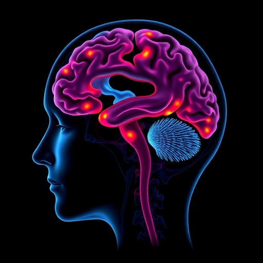
In the relentless pursuit of advancing neurosurgical precision, a groundbreaking study published in Nature Communications outlines how artificial intelligence (AI) is revolutionizing decision-making workflows during the biopsy of diffuse midline gliomas (DMGs), one of the most challenging brain tumors to diagnose and treat. Led by Yuan, Y., Yue, Q., and Wang, Y., among others, this 2025 study introduces a novel intraoperative system leveraging AI to augment real-time pathological assessment of cryosection samples, redefining the biopsy landscape in neurosurgery.
Diffuse midline gliomas are a particularly aggressive class of brain tumors predominantly affecting children and young adults, characterized by a diffuse infiltrative growth pattern located in critical midline structures such as the brainstem and thalamus. Traditional biopsy methods for these tumors present profound clinical challenges—not only because of their precarious anatomical locations but also due to their heterogeneous histopathological features, which demand rapid and accurate intraoperative pathological evaluation. Misdiagnosis or delays in pathological confirmation can have dire consequences on subsequent treatment strategies, emphasizing an urgent need for technological innovation to aid surgical decision-making.
The AI-augmented workflow proposed by Yuan et al. integrates state-of-the-art machine learning algorithms with high-resolution imaging of cryosectioned tumor samples. Cryosection pathology, a rapid tissue freezing and slicing technique, remains a cornerstone in intraoperative diagnostics, providing surgeons with immediate histological insight. However, traditional manual interpretation of these sections by pathologists can be time-consuming, subjective, and prone to error, particularly in complex tumor entities like DMGs. The AI system developed in this study automatically processes and analyzes digital scans of cryosections, identifying malignant features with exceptional accuracy and speed, thus offering robust real-time support during surgery.
Central to the innovation is a deep convolutional neural network (CNN) trained on an expansive dataset of histological images annotated by expert neuropathologists. This AI model is calibrated to discern subtle microscopic patterns that typify diffuse midline gliomas, such as cellular morphology, atypia, and tumor infiltration extent, which may elude even seasoned clinicians under tight operative timelines. Importantly, the algorithm provides quantifiable metrics and visual overlays highlighting key pathological markers, effectively acting as a ‘second pair of eyes’ that enhances diagnostic confidence without supplanting human expertise.
The intraoperative AI system is seamlessly embedded within the surgical workflow. Upon cryosectioning and digitization, the histological images are instantaneously transmitted to a processing unit where the AI model performs rapid segmentation and classification tasks. The results are then relayed back to the surgeon and on-site pathologist through an intuitive interface displaying annotated images alongside predictive analytics. This connectivity ensures that critical histopathological information is accessible within minutes, empowering operative teams to make informed decisions about biopsy adequacy and sampling strategy dynamically.
One of the crucial benefits underscored in this study is the reduction in time from tissue acquisition to diagnosis—a critical parameter in neurosurgery where every second counts. By automating the histological evaluation, the AI-assisted workflow cuts down diagnostic delay significantly compared to conventional manual assessment alone. This expedited turnaround facilitates immediate surgical planning adjustments, potentially improving the extent of tumor sampling and minimizing repeat procedures, which can elevate patient risk.
Yuan and colleagues also demonstrate how AI integration mitigates interobserver variability, a well-documented limitation in neuropathology. Through standardized, reproducible image analysis, the system ensures consistent interpretation of complex biopsy specimens regardless of institutional or individual expertise differences. This standardization is pivotal, especially in centers lacking subspecialized neuropathology services, thus democratizing high-quality diagnostic support for diffuse midline glioma across diverse clinical settings.
The study thoroughly validates the AI model across multiple independent datasets, encompassing variations in staining protocols, imaging equipment, and tumor heterogeneity. The robustness and generalizability of the algorithm affirm its potential for widespread clinical adoption. Importantly, the researchers also emphasize that the AI serves as an adjunct to expert pathologists rather than replacement, promoting synergistic interactions that capitalize on the strengths of both human judgment and machine precision.
Beyond immediate histological classification, the AI system also extracts ancillary features predictive of molecular subtypes and prognostic indicators intrinsic to DMG biology. Integrating such molecular inference during surgery could foreseeably inform personalized therapeutic approaches, marking a paradigm shift towards precision oncology in real time. This capability aligns with burgeoning efforts in cancer research to couple morphological assessment with genomic characterization for superior clinical outcomes.
Ethical considerations and regulatory compliance are resolutely addressed in the manuscript. The authors delineate measures to ensure patient data privacy, algorithm transparency, and rigorous ongoing validation post-deployment. By fostering clinician input throughout developmental phases, the study champions human-centered AI design principles essential for trust and acceptance in surgical practice.
Looking ahead, the authors propose expanding the AI framework to encompass other challenging neuropathological entities and incorporating multi-modal imaging data, such as intraoperative MRI or optical coherence tomography, to enrich diagnostic granularity. The convergence of these technologies holds tremendous implications not only for neurosurgery but broadly across oncologic surgery and pathology disciplines.
The transformative promise of artificial intelligence in the operating theater is exemplified vividly by this pioneering work. By augmenting intraoperative workflows with AI-driven cryosection pathology analysis, Yuan, Yue, Wang, and their team have illuminated a path towards more precise, rapid, and standardized biopsy procedures in diffuse midline glioma management. This innovation stands to significantly impact patient prognosis by enabling tailored strategies promptly, reducing surgical risks, and optimizing therapeutic efficacy in battling one of the most formidable brain cancers.
As AI continues its inexorable integration into medicine, the partnership between machine intelligence and human expertise forged here serves as a compelling model for future clinical innovations. Harnessing the synergy of computational power and clinical acumen, this study heralds a new era in neurosurgical oncology where decisions are not only swifter but smarter and more individualized. The ripple effects of such advancements promise to outlast the operating room, fundamentally reshaping how complex cancers are understood and treated.
Ultimately, this research is more than an incremental technical achievement—it is a bold leap forward, charting a course toward actionable AI in the very heart of surgical care. It challenges the medical community to rethink workflows, embrace emerging technologies, and prioritize patient-centered outcomes powered by data-driven insights. The integration of AI into biopsy pathology for diffuse midline gliomas is poised to redefine what is possible in neuro-oncology, offering renewed hope for improved survival and quality of life in a domain previously marked by diagnostic uncertainty and therapeutic limitations.
Subject of Research: Artificial intelligence-augmented intraoperative workflows for biopsy pathology in diffuse midline glioma.
Article Title: AI-augmented intraoperative decision-making workflows in diffuse midline glioma biopsy using cryosection pathology.
Article References:
Yuan, Y., Yue, Q., Wang, Y. et al. AI-augmented intraoperative decision-making workflows in diffuse midline glioma biopsy using cryosection pathology. Nat Commun (2025). https://doi.org/10.1038/s41467-025-66853-y
Image Credits: AI Generated
Tags: advanced surgical workflowsAI in neurosurgeryartificial intelligence pathology assessmentbrain tumor diagnosis challengescryosection pathology techniquesdiffuse midline glioma biopsyimproving biopsy accuracyintraoperative decision-makingmachine learning in healthcarepediatric brain tumorsreal-time pathological evaluationtechnological innovation in medicine


