Heart development is a fascinating and complex process that has captivated researchers for decades. The heart is not only the first functional organ to form during embryonic development, but it also plays a crucial role in sustaining life by circulating blood throughout the body. Understanding how the heart develops requires exploring its intricate anatomical, cellular, and molecular frameworks. Researchers have dedicated their careers to deciphering the multitude of factors that contribute to cardiogenesis, yet challenges persist in fully appreciating how tissue organization and cellular lineages integrate to form a synchronized and functional heart.
In recent years, advances in imaging technologies and genomics have provided unprecedented insights into heart development. These technological advancements serve as vital tools, allowing researchers to observe the heart’s intricate formation as it evolves from a simple tube to a complex organ with distinct chambers and specialized cell types. The field has seen a shift from traditional observational methods to dynamic, high-resolution imaging that illuminates cellular behaviors in real time, revealing the subtleties of cardiac morphogenesis in ways previously thought impossible.
The heart tube is the first structure to arise during heart development, forming from the mesoderm layer of the developing embryo. This tube undergoes a series of morphogenetic transformations, folding, and looping to create the basic architecture of the heart. Understanding these early stages is essential as they set the foundation for subsequent heart maturation processes. Researchers have identified key molecular signals that guide the formation and remodeling of the heart tube, unraveling the genetic networks that govern this critical phase of development.
Central to heart development is the process of cardiomyocyte differentiation. Cardiomyocytes are the beating cells of the heart, and their proper formation is crucial for establishing a functional cardiac muscle. Recent studies have revealed the roles of various signaling pathways, such as Wnt, Notch, and BMP, in promoting cardiomyocyte lineage specification from progenitor cells. These molecular cues are intricately timed and precisely coordinated to ensure that the heart develops with the correct cellular composition and functional capabilities.
As the heart develops, so does its electrical conduction system. The establishment of the initial heartbeat is a remarkable event, marking the transition from embryonic development to functional organ. This electrophysiological activity is driven by specialized pacemaker cells that emerge during early heart development. Understanding the mechanisms that lead to pacemaker cell formation has been a focal point of research, with implications for treating arrhythmias and other cardiac disorders.
The interplay between morphogenesis and function is another critical aspect of heart development. As the heart matures, its structural integrity is reinforced, and its functional capacities increase. The coordinated growth of cardiac chambers and the vascular system is essential for optimizing blood flow and maintaining homeostasis. Researchers have begun to appreciate how mechanical forces influence heart development, as the heart’s shape and size adjust in response to hemodynamic stress. This feedback loop highlights the importance of environmental factors in the developmental process.
In vitro models, particularly cardiac organoids, have emerged as revolutionary tools for studying heart development. These three-dimensional organ-like structures can recapitulate some aspects of cardiogenesis, allowing scientists to observe cellular interactions and tissue organization in a controlled environment. By utilizing stem-cell-derived cardiomyocytes, researchers can investigate how different cellular components contribute to heart function and development. The insights gained from these organoid models have the potential to transform our understanding of congenital heart defects and provide avenues for novel therapeutic strategies.
The exploration of cardiac development is further enriched by the integration of omics technologies, including genomics, transcriptomics, and proteomics. These high-throughput approaches provide a systems-level view of heart development, enabling researchers to identify gene expression patterns, signaling networks, and molecular interactions involved in cardiogenesis. Such comprehensive datasets can highlight key regulators of heart development and identify potential targets for therapeutic intervention in heart disease.
The implications of studying heart development extend beyond basic science. Understanding the mechanisms that govern cardiogenesis may pave the way for innovative treatments for congenital heart defects, which are among the most common types of birth defects globally. By dissecting the underlying causes of these conditions, researchers can devise strategies to repair or regenerate damaged cardiac tissue, offering hope to millions of patients.
Current studies are increasingly focusing on the intersection of genetics and environment in heart development. The influence of epigenetic modifications, environmental exposures, and maternal health on cardiogenesis is an exciting frontier in cardiac research. Insights into how these factors interact with intrinsic genetic programming could provide a holistic understanding of heart development and disease susceptibility, leading to more personalized approaches in medicine.
As the field advances, ethical considerations surrounding the use of stem cells and organoids in research continue to spark discussions. The potential for creating heart tissues for transplantation or modeling disease raises questions about the moral implications of manipulating life at the cellular level. Striking a balance between scientific progress and ethical responsibility will be crucial as researchers navigate the rapidly evolving landscape of cardiac development studies.
In summary, the multi-faceted exploration of heart development encompasses a wide range of approaches, from high-resolution imaging to advanced in vitro models. A comprehensive understanding of how the heart forms and functions is essential not only for basic science but also for developing therapeutic strategies for heart-related diseases. The ongoing research efforts hold great promise for unraveling the complexities of cardiogenesis and translating these discoveries into meaningful clinical applications.
We stand at the cusp of a new era in cardiovascular research, armed with powerful tools and technologies that have the potential to transform our comprehension of heart development. As researchers continue to forge ahead, the future looks bright for unraveling the mysteries of the heart and enhancing our understanding of its development and function.
Subject of Research: Heart Development
Article Title: Coordination of cardiogenesis in vivo and in vitro
Article References:
Mendjan, S., Deyett, A. & Yelon, D. Coordination of cardiogenesis in vivo and in vitro.
Nat Rev Mol Cell Biol (2025). https://doi.org/10.1038/s41580-025-00878-5
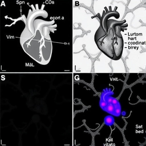
Image Credits: AI Generated
DOI:
Keywords: Cardiogenesis, Heart Development, Stem Cells, Cardiac Organoids, Electrophysiology, Molecular Signaling, Congenital Heart Defects.
Tags: advances in genomics for heart researchcardiac tissue organizationcellular and molecular frameworkschallenges in heart development researchdynamic imaging technologiesembryonic cardiogenesisheart development processheart morphogenesis imagingheart structure formationin vitro heart modeling techniquesin vivo heart development studiessynchronized heart function integration


