A New Era in Brain Mapping: Introducing NextBrain, a Probabilistic Histological Atlas Revolutionizing MRI Segmentation
The neuroscience community stands on the brink of a transformative breakthrough in brain mapping with the introduction of NextBrain, a cutting-edge probabilistic human brain atlas engineered for unparalleled MRI segmentation. Unlike traditional brain atlases that offer static representations, NextBrain integrates probabilistic models with high-resolution imaging data, creating a dynamic, versatile tool tailored for both research and clinical applications. This innovation, unveiled in a recent study, represents not just an incremental advance but a fundamental leap forward in our capacity to visualize, analyze, and interpret human brain architecture at unprecedented granularity.
NextBrain emerges as a comprehensive resource, freely accessible to researchers worldwide. Its foundation is a meticulously curated multimodal dataset encompassing raw and registered ex vivo brain MRI scans at a remarkable 100-micrometer resolution. This level of detail unlocks new potentials for population-level analyses, allowing subtle neuroanatomical variations across individuals to be quantified with enhanced precision. The dataset’s openness invites global collaboration, enabling scientists to repurpose, augment, and extend these foundational data—whether by refining region-of-interest (ROI) delineations using histological or MRI inputs, or by constructing bespoke atlases tailored to specific research queries.
Of particular note is NextBrain’s robust Bayesian segmentation tool, a sophisticated algorithmic framework that leverages prior probabilistic knowledge to distinguish and segment brain regions automatically. This approach not only harnesses the power of next-generation imaging but also adapts seamlessly to diverse scanning conditions, including the challenging interface between ex vivo and in vivo data. Its compatibility with standard 1-mm isotropic scans ensures broad applicability, from high-field laboratory MRI systems to routine clinical imaging environments. By marrying rich probabilistic models with real-world imaging constraints, NextBrain sets a new standard for accurate, reproducible, and granular brain segmentation.
The atlas itself features a high-resolution Common Coordinate Framework (CCF) designed for population analyses, offering a scaffold for integrating diverse neuroimaging data. This CCF promotes interoperability and serves as a pivotal reference for cross-study comparisons, thus facilitating meta-analyses and longitudinal studies. Moreover, the ex vivo MRI scans embedded within the dataset afford an invaluable complement to conventional in vivo imaging, capturing microscale anatomical details that were previously inaccessible and thereby filling critical gaps in our understanding of brain structure and connectivity.
NextBrain’s design emphasizes customizability and extensibility, reflecting the modern paradigm of open science and collaborative innovation. Researchers are empowered to download the full dataset and codebase, modify annotations, or incorporate novel brain regions according to their study needs. This modular architecture invites the integration of other state-of-the-art segmentation methods and atlases, including advances in cortical surface modeling. The team behind NextBrain is actively developing hybrid models that blend neural networks with geometric processing techniques to extract laminar-specific segmentations—an ambitious endeavor aimed at resolving the fine-scale layering of the cortex from both ex vivo and in vivo scans.
One of the key visionary aspects of NextBrain is its adaptability to emerging imaging modalities. As ultra-high-field MRI scanners (such as 7 Tesla systems) and novel technologies like Hierarchical Phase-Contrast Tomography (HiP-CT) become more accessible, the atlas can fully leverage these high-resolution datasets. This ensures that NextBrain remains at the forefront of neuroimaging innovation, capable of supporting the next wave of discoveries in brain microstructure and pathology by capitalizing on ever-improving imaging fidelity and contrast mechanisms.
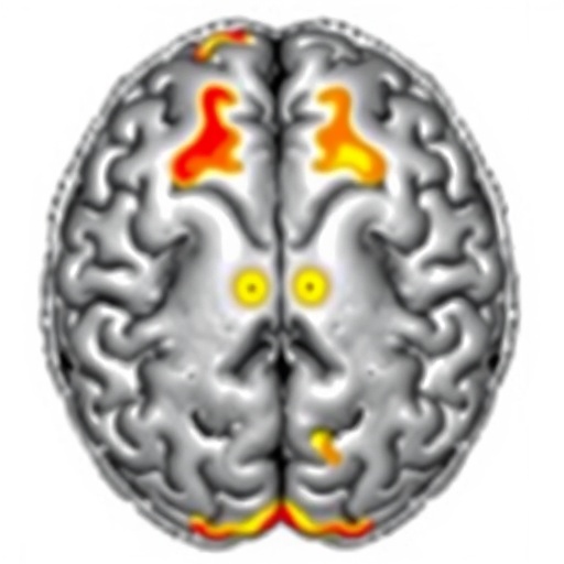
Beyond healthy brain anatomy, NextBrain’s probabilistic framework holds transformative promise for studying neurological diseases. The incorporation of pathology models within the Bayesian segmentation pipeline enables automated delineation of abnormal tissue, such as tumours or white matter hyperintensities, often linked to neurodegenerative conditions and cerebrovascular disease. This capacity paves the way for objective and detailed quantification of disease burden, which is critical for advancing diagnostic criteria, monitoring progression, and evaluating therapeutic interventions in diseases like Alzheimer’s and multiple sclerosis.
The development of NextBrain underscores an exciting shift towards integrative, data-driven neuroscience, where anatomy, pathology, and function can be mapped with unprecedented consistency and resolution. By providing a common language and probabilistic foundation, this atlas facilitates the synthesis of diverse datasets across institutions and modalities, accelerating the pace of discovery. It also democratizes access to state-of-the-art brain mapping tools, empowering researchers with varied expertise and resources to contribute meaningfully to the neuroscience frontier.
NextBrain’s Bayesian segmentation approach also offers significant improvements in reproducibility and accuracy over traditional deterministic segmentation pipelines. By explicitly modeling uncertainty and variability within anatomical structures, the approach yields more robust outputs, especially when dealing with noisy or artefact-prone images—common challenges in clinical neuroimaging. This probabilistic dimension enhances confidence in downstream analyses, from volumetric assessments to connectivity mapping, making NextBrain a critical asset for both basic research and translational neuroscience.
The underlying architecture of NextBrain enables seamless augmentation as new anatomical regions are characterized. The ability to iteratively update and refine ROIs based on histology or advanced MRI contrasts means the atlas evolves alongside scientific knowledge. This forward-compatible design ensures NextBrain remains a living resource rather than a fixed repository, stimulating continual improvements driven by community engagement and technological advances.
Importantly, the open-access ethos of NextBrain fosters widespread adoption and interdisciplinary collaboration. By removing barriers to high-quality brain atlases and segmentation tools, the project catalyzes innovation beyond traditional neuroimaging labs, opening opportunities in computational neuroscience, neuroinformatics, and even clinical neurology. This broad dissemination could hasten breakthroughs in understanding brain development, aging, and disease progression, ultimately impacting patient care and therapeutic development on a global scale.
In conclusion, NextBrain epitomizes the future of brain atlas construction and MRI segmentation, merging probabilistic modeling, high-resolution multimodal data, and open-source accessibility into a powerful, extensible platform. Its introduction heralds a new era where detailed, personalized, and accurate brain maps are within reach for researchers and clinicians alike. As imaging technologies continue to evolve and datasets expand, NextBrain’s flexible framework ensures that the neuroscience community is equipped to extract ever richer insights into the human brain’s complex organization and its disorders, paving the way for revolutionary advances in brain science.
Subject of Research: Probabilistic human brain atlas and MRI segmentation methods
Article Title: A probabilistic histological atlas of the human brain for MRI segmentation
Article References:
Casamitjana, A., Mancini, M., Robinson, E. et al. A probabilistic histological atlas of the human brain for MRI segmentation. Nature (2025). https://doi.org/10.1038/s41586-025-09708-2
Image Credits: AI Generated
DOI: https://doi.org/10.1038/s41586-025-09708-2
Keywords: Brain atlas, MRI segmentation, probabilistic modeling, Bayesian segmentation, ex vivo MRI, ultra-high-field MRI, HiP-CT, neuroimaging, neuroanatomy, open-access dataset, cortical parcellation, neuropathology
Tags: Bayesian modeling in brain researchclinical applications of brain atlasescollaborative brain research initiativesdynamic brain mapping techniqueshigh-resolution brain MRI scansMRI segmentation innovationmultimodal brain imaging dataneuroanatomical variation quantificationNextBrain neuroscience toolopen-access neuroscience resourcespopulation-level neuroanatomical analysisProbabilistic brain atlas

