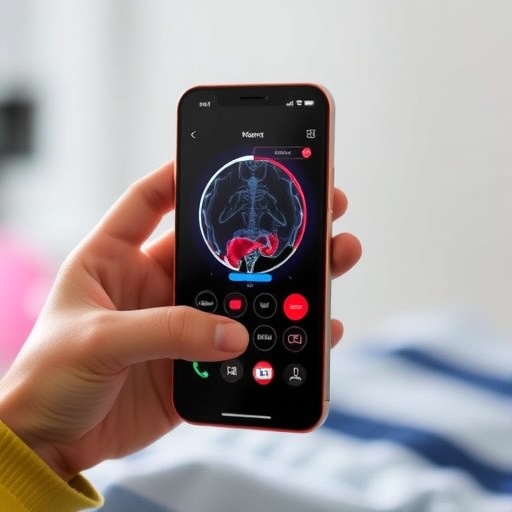
In a groundbreaking advance for neuromuscular disease diagnostics and treatment monitoring, researchers from Stanford University have demonstrated that simple smartphone cameras can replace traditional stopwatch methods and even rival sophisticated, high-cost motion laboratories. This innovative approach was detailed in a study published in the New England Journal of Medicine AI, showcasing how video-based biomechanical analysis can capture nuanced disease-specific movement signatures with unprecedented precision.
For decades, clinicians relied on timed function tests, often measured with nothing more than a stopwatch, to evaluate patients with neuromuscular conditions such as facioscapulohumeral muscular dystrophy (FSHD) and myotonic dystrophy (DM). These tests, while quick and inexpensive, provide only a surface-level understanding of patient mobility, failing to detect subtle biomechanical changes indicative of disease progression or response to therapy. The new research addresses this limitation by harnessing smartphone video and advanced computational modeling to quantitatively analyze movement with laboratory-level accuracy.
At the core of this innovation is OpenCap, an open-source software platform developed by the Stanford team. Utilizing footage from up to three synchronized smartphone cameras, OpenCap reconstructs a three-dimensional digital twin of the patient’s biomechanics as they perform a series of clinical movements. These include tasks such as walking ten meters, running, and raising calves. The system extracts over thirty movement metrics relevant to neuromuscular function, including stride length, range of motion, joint angles, and gait kinematics, providing a rich dataset far beyond elapsed time measures.
The study involved nearly 130 participants, two-thirds of whom were diagnosed with either FSHD or DM. By processing videos of these individuals performing nine specific movements, researchers demonstrated that the smartphone-derived timing metrics strongly correlated with traditional stopwatch measurements, exhibiting comparable reliability upon test repetition. More importantly, the video data unveiled subtle but distinct biomechanical patterns unique to each disease, such as shorter strides coupled with higher ankle elevation in FSHD, or difficulty rising from a chair in DM patients, which conventional timed tests failed to detect.
Leveraging machine learning classifiers on these movement features, the team achieved an impressive 82% accuracy in identifying a patient’s specific neuromuscular disease, significantly outperforming standard stopwatch-based diagnosis accuracy, which hovered near chance at 50%. This signals a paradigm shift where remote, rapid, and automated biomechanical assessments can contribute not only to disease monitoring but potentially to early diagnosis.
The implications for clinical trials are profound. High-fidelity motion capture traditionally required expensive equipment and specialists, limiting assessments to sporadic, resource-intensive sessions in motion labs. OpenCap democratizes access by enabling easy, cost-effective data collection anywhere with just a smartphone. This technological leap allows for more frequent, objective, and detailed monitoring of disease progression or therapeutic response, which could accelerate drug development and personalized treatment strategies.
Stanford bioengineering professor Scott Delp, the senior author on the study, emphasized how integrating sophisticated biomechanical modeling with ubiquitous smartphone hardware bridges the gap between experimental research and everyday clinical practice. The ability to generate digital biomechanical twins paves the way for real-time feedback and more nuanced functional assessments, aligning diagnostics with the molecular precision emerging in pharmacological therapies.
Beyond neuromuscular diseases, OpenCap is already being utilized globally in diverse applications, including sports medicine. For example, Germany’s national volleyball team employed the technology to evaluate injury risk and optimize athlete performance, condensing what once took years of data collection into a single season. This demonstrates the platform’s versatility and potential to revolutionize human movement analysis across disciplines.
Despite the technology’s promise, Delp and colleagues caution that ongoing validation and adaptation are necessary to ensure accuracy across different patient populations and clinical settings. Future research will focus on refining algorithms, expanding disease coverage, and integrating these tools seamlessly into clinical workflows and electronic health records. The ultimate vision is a future where comprehensive biomechanical assessment is as accessible as a routine vital sign.
In effect, this study heralds a future where healthcare professionals can leverage everyday devices to perform detailed functional evaluations, enhance diagnostic precision, and personalize treatment regimens on an unprecedented scale. As neuromuscular disease therapies continue to evolve, such innovations will be pivotal in detecting early improvements or setbacks, empowering clinicians and patients alike.
The convergence of mobile technology, computer vision, and biomechanics marks a turning point in how movement disorders are understood and managed. It unlocks a new era in digital health, where scalable, portable, and sophisticated tools are no longer confined to specialized centers but available in clinics, homes, and communities worldwide. The potential impact on patient outcomes and healthcare delivery is both tangible and transformative.
With continuing advances and widespread adoption, smartphone-based biomechanical analysis could soon become a standard tool in neurology and rehabilitation medicine. By democratizing access to detailed movement data, this approach promises to accelerate research, improve clinical decision-making, and ultimately enhance quality of life for millions afflicted with neuromuscular diseases.
Subject of Research: People
Article Title: Video-Based Biomechanical Analysis Captures Disease-Specific Movement Signatures of Different Neuromuscular Diseases
News Publication Date: 28-Aug-2025
Web References: http://dx.doi.org/10.1056/AIoa2401137
Keywords: Muscular dystrophy, Bioengineering
Tags: advanced movement analysis for FSHDAI in medical diagnosticsbiomechanical analysis with smartphonescomputational modeling in medicinedigital twins in patient monitoringmonitoring neuromuscular diseasesOpenCap software for biomechanicspatient mobility assessment innovationsprecision medicine in neuromuscular disorderssmartphone cameras in clinical evaluationsmartphone technology in healthcarevideo-based diagnostics for myotonic dystrophy

