In an age where precision medicine is becoming increasingly vital, researchers are exploring innovative methodologies to enhance the understanding of human anatomy. A groundbreaking study led by Duquesne et al. meticulously investigates the complex geometry of the human hand and foot skeleton using advanced statistical shape modeling techniques. This work, published in the “Annals of Biomedical Engineering,” aims to bridge the gap between external anatomical features and internal skeletal structures, unlocking new possibilities for clinical applications ranging from prosthetics to orthopedic interventions.
The study meticulously develops a robust statistical framework to predict the geometric configuration of the bony structures of hands and feet. By employing sophisticated algorithms, the researchers were able to analyze a vast dataset of anatomical scans, allowing them to create detailed models of skeletal geometry. The use of statistical shape modeling (SSM) techniques enables a comprehensive understanding of shape variance, providing insights into how individual anatomy can differ even within a seemingly uniform population. This is particularly relevant in fields such as personalized medicine, where tailored interventions could substantially enhance patient outcomes.
This research contributes to a wider understanding of how anatomical diversity affects clinical practices. Standard models of hand and foot anatomy often overlook significant variations found in different populations. By focusing on the skeletal topology, the authors aim to improve anthropometric data that is essential for the design of medical devices, including customized implants and orthoses. This shift toward individualized designs can be fundamental in addressing the intricate requirements of patients who exhibit unique anatomical features.
The foundation of this study lies in a well-defined methodological approach that involves data acquisition, shape modeling, and statistical analysis. The researchers employed a diverse cohort for their data collection, ensuring that the resulting models reflect a wide range of anatomical variations. By integrating machine learning with traditional shape analysis, the team has pioneered a method that not only captures complex shapes but also predicts the likelihood of specific anatomical configurations. This dual approach paves the way for more dynamic and flexible modeling techniques that can be applied across various applications in medical science.
One of the key advantages of utilizing statistical shape models is the ability to visualize and comprehend complex shape interactions better. For the hand and foot, where bone structure intricately relates to functional movement, understanding these geometric nuances is essential. The statistical framework employed in this research results in a three-dimensional representation of bony structures that can aid surgeons and clinicians in pre-operative planning. Moreover, these models can help in identifying potential issues prior to surgical interventions, ultimately leading to safer and more effective procedures.
In conjunction with the technical advancements, the study also emphasizes the significance of collaboration between engineering and medical fields. By fostering interdisciplinary partnerships, researchers can harness the power of computational modeling, machine learning, and clinical insight to enhance healthcare delivery. This integrated approach is particularly relevant in the context of developing adaptive prosthetic devices that mimic natural movement patterns. By closely aligning device design with anatomical insights, engineers can create solutions that significantly improve the quality of life for individuals with limb differences.
Furthermore, the implications of this research extend beyond clinical realms into educational settings. By understanding the skeletal variations and their implications, educators can better prepare future medical professionals to address the complexities of human anatomy. This study serves as a stepping stone for future research initiatives aimed at enhancing anatomical education, ensuring that upcoming generations of healthcare practitioners possess an in-depth understanding of human variability.
Despite the promising insights gained from this research, the authors acknowledge the limitations inherent in their study. As with any statistical model, there exists a certain level of unpredictability when applying these findings to individual cases. Future work will focus on refining these models to account for additional variables such as age, gender, and health status, which can all substantially influence skeletal geometry. By continuously updating and validating these models against real-world data, researchers can enhance their reliability and applicability in clinical settings.
The exploration of bony geometry through statistical shape modeling also opens avenues for clinical research focused on various musculoskeletal disorders. Conditions such as arthritis, osteoporosis, and congenital anomalies can profoundly affect skeletal structure and function. By leveraging the methods developed in this study, clinicians could develop predictive models that identify at-risk individuals earlier in their treatment journey. This proactive approach could transform patient care, enabling timely interventions and personalized therapy plans based on predictive analytics.
In summary, the work by Duquesne et al. signifies a pivotal shift in how medical and engineering fields can converge to tackle challenges associated with anatomical variability. Through statistical shape modeling, researchers have not only advanced our understanding of skeletal geometry but have also laid the groundwork for future innovations in personalized medicine, surgical planning, and educational practices. As the landscape of healthcare continues to evolve, integrating these sophisticated modeling techniques will be crucial in optimizing patient care.
Ultimately, this research represents a vibrant intersection of technology and biology, showcasing how interdisciplinary collaboration can lead to unprecedented advancements in understanding human anatomy and improving health outcomes. By embracing this innovative approach, the medical community can expect significant breakthroughs in not just how we treat skeletal issues but also in fostering a comprehensive understanding of the complex interplay between form and function within the human body.
As the study unfolds and its insights are further explored, the potential for translating these findings into clinical practice seems boundless. The emphasis on precision and individualization aligns flawlessly with the trend towards tailored healthcare solutions, making statistical shape modeling a promising frontier in biomedical engineering.
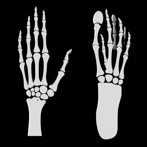
Subject of Research: Statistical Shape Modeling for Hand and Foot Bony Geometry
Article Title: From Skin to Skeleton: A Statistical Shape Modelling Approach for Predicting Hand and Foot Bony Geometry
Article References:
Duquesne, K., Molnar, A., Huysentruyt, R. et al. From Skin to Skeleton: A Statistical Shape Modelling Approach for Predicting Hand and Foot Bony Geometry. Ann Biomed Eng (2025). https://doi.org/10.1007/s10439-025-03882-0
Image Credits: AI Generated
DOI: https://doi.org/10.1007/s10439-025-03882-0
Keywords: Statistical Shape Modeling, Bony Geometry, Hand, Foot, Personalized Medicine, Clinical Applications, Biomedical Engineering.
Tags: advanced statistical analysis in medicineanatomical diversity in clinical practiceanatomical scans dataset analysisbridging external features and internal structuresclinical applications of skeletal modelinggeometric modeling of skeletal structureshuman hand and foot anatomypersonalized medicine applicationsprosthetics and orthopedic interventionsrobust statistical frameworks in researchstatistical shape modeling techniquesunderstanding bone shape variance

