Title: Evaluating the Reliability of Cone Beam CT in Treating Class III Growing Patients: A Deep Dive into Craniomaxillary Changes
Recent advancements in orthodontics have brought cone beam computed tomography (CBCT) to the forefront of diagnostic imaging, particularly for assessing craniofacial structures. A pivotal study conducted by Hu, Zhang, Cheung, and their colleagues investigates the reliability and reproducibility of CBCT assessments concerning craniomaxillary changes before and after treatment in Class III growing patients. This approach not only highlights the effectiveness of CBCT technology but also emphasizes the necessity for precise treatment methodologies.
CBCT technology has revolutionized how orthodontists visualize and analyze craniofacial abnormalities. The ability to obtain three-dimensional images yields invaluable information that traditional two-dimensional radiographs often overlook. However, the reliability of such imaging techniques is crucial, particularly in young patients whose maxillofacial structures are still developing. The focus of the current study seeks to establish a critical evaluation of how CBCT can be reliably employed in clinical practice, specifically for Class III malocclusion.
Class III malocclusion, characterized by the mandibular dental arch being positioned farther forward than the maxillary arch, presents unique challenges in diagnosis and treatment. Understanding the underlying craniofacial alterations is crucial for orthodontists to develop effective treatment plans. This study provides a foundation for discerning whether the data derived from CBCT scans can be trusted to predict treatment outcomes accurately. Moreover, ensuring the reproducibility of these measurements establishes a clear pathway for consistent orthodontic practices.
The research comprised a comprehensive analysis of various factors influencing the reliability of CBCT assessments. It scrutinizes how different imaging methods can yield varied results, prompting practitioners to exercise caution in their interpretations. Indeed, the study generates an essential dialogue on the responsibilities of both orthodontists and radiologists in ensuring that the data from CBCT scans are accurate, reliable, and ultimately beneficial for the patients.
Furthermore, the study outlines the experimental methodologies adopted by the researchers, who employed a cohort of Class III patients undergoing orthodontic treatment. Using dual CBCT scans — one taken before treatment initiation and another captured post-treatment — the researchers detected significant morphological changes. Notably, these changes were meticulously quantified using reliable measurements, ensuring that conclusions are both meticulous and grounded in evidenced data.
Reliability in imaging not only hinges on the technology itself but also on the technician’s expertise and the methodology chosen. The researchers emphasized that protocols used during scanning significantly impact the quality of imaging outcomes. For instance, patient positioning and scan parameters must be meticulously managed to minimize artifacts that could distort the resultant images. This insight presents an opportunity for significant improvements within clinical settings by developing standard operating procedures that can enhance the imaging quality of CBCT scans.
Moreover, the researchers found that one of the primary benefits of employing CBCT lies in its capacity to visualize not only the bony structures but also the soft tissue contours. This comprehensive imaging capability offers orthodontists detailed insight into craniofacial dynamics, enhancing their ability to assess individual patient needs accurately. This study underscores the importance of integrated diagnostic tools in providing personalized treatment plans for Class III patients.
While the advancement in imaging technologies is significant, the critical aspect of this study lies in its call for caution. The authors consistently highlight that while the capabilities of CBCT are impressive, they come with inherent limitations that specialists must acknowledge. Potential pitfalls include reliance on complex algorithms and software that could inadvertently lead to discrepancies in diagnoses if not used correctly.
The researchers also propose that future studies should focus on longitudinal assessments to ascertain the long-term reliability of CBCT evaluations in various treatment stages. Such data will be essential for validating the initial findings. By establishing long-term follow-ups, orthodontists can better understand how craniomaxillary changes evolve over years, contributing significantly to future clinical practices.
In conclusion, the reliance and reproducibility of CBCT assessments of craniomaxillary changes represent a crucial area of research within orthodontics, particularly concerning Class III growing patients. This study by Hu and colleagues lays the groundwork for developing enhanced imaging protocols while also advocating for prudent utilization of CBCT technology in clinical settings. As the field of orthodontics continues to embrace technological advancements, commitment to accuracy and reliability remains paramount for delivering effective orthodontic care.
As the field progresses, embracing a cautious or critical approach may ultimately lead to improved patient outcomes and more effective bracing solutions, ensuring that every Class III patient receives the highest standard of treatment tailored to their unique anatomical needs.
Subject of Research: The reliability and reproducibility of CBCT assessment in orthodontics for craniomaxillary changes.
Article Title: Reliability and reproducibility of CBCT assessment of craniomaxillary changes before and after treatment for Class III growing patients – a cautious or critical approach.
Article References:
Hu, X., Zhang, Y., Cheung, G.S.P. et al. Reliability and reproducibility of CBCT assessment of craniomaxillary changes before and after treatment for Class III growing patients – a cautious or critical approach.
BMC Pediatr 25, 794 (2025). https://doi.org/10.1186/s12887-025-06049-x
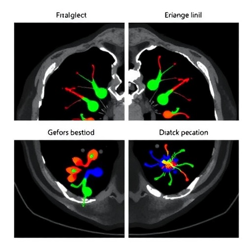
Image Credits: AI Generated
DOI: 10.1186/s12887-025-06049-x
Keywords: CBCT, Class III malocclusion, orthodontics, craniomaxillary changes, reliability, reproducibility, imaging technology.
Tags: CBCT reliability in orthodonticsClass III malocclusion treatmentclinical evaluation of CBCTcone beam CT technology benefitscraniomaxillary changes imaginggrowing patients orthodonticsmaxillofacial development assessmentorthodontic diagnostic advancementsorthodontic imaging techniquesorthodontic treatment methodologiesorthodontic treatment planning for Class III patientsthree-dimensional imaging in dentistry


