In the ever-evolving world of cellular biology, precise visualization and measurement of cell structures are paramount to decoding the myriad functions cells perform. Recent advancements from researchers at the University of Tsukuba in Japan introduce DeMemSeg, an AI-powered, deep learning-based pipeline markedly enhancing the segmentation of overlapping cell membranes derived from two-dimensional projections of three-dimensional fluorescence images. This breakthrough represents a significant leap in automating and refining morphological analyses of individual cellular components, particularly within the study of budding yeast.
Cell morphology, particularly the architecture and dynamics of cell membranes and organelles, is intricately linked to cellular function. The challenge in biological imaging lies in the accurate translation of complex three-dimensional (3D) structures into two-dimensional (2D) formats—especially 2D projections derived from fluorescence microscopy images commonly used in biological research. This dimensional reduction often results in the visual overlap of distinctly separated membranes and organelles, complicating efforts to delineate individual cells or subcellular structures with precision.
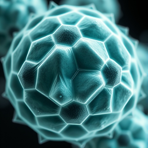
The DeMemSeg pipeline spearheads a novel approach to this conundrum. By leveraging a customized deep learning framework, this method excels at parsing overlapping membrane segments in 2D projections, which historically has been a prohibitive challenge in image-based morphological analysis. The pipeline was developed using the prospore membrane—a specialized membrane formed during the sporulation phase in budding yeast—as its experimental model, a system well-suited for probing the nuances of membrane morphology and dynamics.
Validation of DeMemSeg was rigorous, with the algorithm’s accuracy benchmarked against expert manual segmentation, often regarded as the gold standard despite being labor-intensive and subjective. Remarkably, the AI-driven method demonstrated statistically comparable performance, signaling its readiness for widespread adoption. Moreover, the AI’s robustness was tested against mutant yeast strains exhibiting abnormal membrane morphologies absent from the original training dataset. Here, DeMemSeg not only detected these morphological aberrations but quantified them effectively, illustrating an impressive capacity for generalization beyond its initial training scope.
This AI pipeline’s capacity to dissect complex overlapping features in 2D projections heralds a paradigm shift in high-throughput morphological analyses. Traditional methods have struggled with scalability and throughput owing to manual intervention or simplistic segmentation algorithms. DeMemSeg offers a scalable, objective, and reproducible solution capable of transforming large-volume data into interpretable quantitative insights, which is particularly valuable in research involving budding yeast, a model organism pivotal for understanding fundamental biological processes.
Beyond its application in yeast biology, the implications of DeMemSeg’s technology are profound and far-reaching. Morphological irregularities in membranes and organelles are hallmarks of several human disorders related to gamete formation and fertilization. By providing a robust tool for the high-resolution characterization of cell membrane morphology, this AI-driven pipeline lays the groundwork for elucidating complex disease mechanisms, potentially accelerating diagnostics and therapeutic interventions in reproductive biology and beyond.
The technical sophistication of DeMemSeg lies in its underlying deep learning architecture. Unlike conventional image segmentation techniques reliant on fixed algorithms or thresholding, this AI model dynamically adapts to diverse and noisy imaging data, leveraging convolutional neural networks trained on meticulously curated datasets. This adaptability enables the pipeline to maintain accuracy even when confronted with novel morphological phenotypes or imaging artifacts, addressing a critical bottleneck in biomedical image analysis.
Furthermore, DeMemSeg integrates seamlessly into the research workflow, automating the generation of high-quality, cell-level training data sets that can be iteratively refined and expanded. This feature not only accelerates initial development but also facilitates continuous learning and adaptation as new data types or experimental conditions emerge, ensuring long-term utility in various investigative contexts.
The success of DeMemSeg in accurately segmenting overlapping membranes in fluorescence microscopy projections also opens avenues for integration with other imaging modalities. Combining this AI-driven segmentation with high-resolution live-cell imaging, multi-spectral fluorescence techniques, or even volumetric imaging approaches could vastly enhance the temporal and spatial understanding of membrane dynamics, cellular interactions, and developmental processes in complex biological systems.
With the burgeoning availability of large-scale biological image repositories and advances in AI, tools like DeMemSeg epitomize the next frontier in systems biology and computational microscopy. By automating the laborious process of cell segmentation with unprecedented precision and scalability, the research community gains a powerful asset to unravel cellular heterogeneity, morphological variability, and pathological transformations more effectively than ever before.
This convergence of artificial intelligence, imaging technology, and cellular biology exemplifies the transformative potential of interdisciplinary research. The resulting insights not only propel the foundational understanding of life sciences but also inform translational research efforts targeting human health and disease. As DeMemSeg continues to evolve, its application may extend to diverse fields such as developmental biology, neurobiology, and cancer research—domains where membrane morphology and cell architecture are equally critical.
In summary, DeMemSeg represents a significant advancement in quantitative cell biology, offering a compelling solution to the longstanding challenge of accurately segmenting overlapping membranes from 2D projections. By combining deep learning algorithms with biological expertise, this pipeline enhances the reliability and throughput of morphological analyses, paving the way for new discoveries in cell function, pathology, and therapeutic development.
Subject of Research: Cell membrane morphology and segmentation in budding yeast using AI-driven deep learning techniques.
Article Title: Deep learning-based segmentation of 2D projection-derived overlapping prospore membrane in yeast
News Publication Date: 13-Sep-2025
Web References: https://doi.org/10.1247/csf.25032
References: Deep Learning-Based Segmentation of 2D Projection-Derived Overlapping Prospore Membrane in Yeast, Cell Structure and Function, DOI: 10.1247/csf.25032
Image Credits: University of Tsukuba
Keywords: Cell biology, Cell morphology, Cell membranes, Budding yeasts, Image analysis, Quantitative analysis, Data visualization, Image segmentation, Artificial intelligence
Tags: 3D to 2D imaging challengesAI-driven segmentation techniquesautomated image processing in biologybudding yeast cellular studiescell morphology visualizationdeep learning in cellular biologyDeMemSeg pipeline developmentfluorescence microscopy advancementsmorphological analysis of cell structuresoverlapping cell membranes analysissubcellular structure delineationUniversity of Tsukuba research breakthroughs


