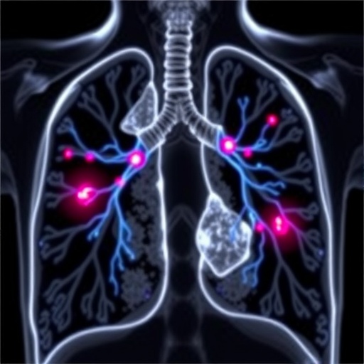
Recent advancements in the field of medical imaging have ushered in a new era of precision medicine, particularly in the diagnosis and classification of various cancers. Among these cutting-edge developments, a groundbreaking study has emerged that explores multi-domain histopathological imaging biomarkers for the classification of two prevalent types of lung cancer: squamous cell carcinoma (SCC) and adenocarcinoma (AC). This research, spearheaded by Liu et al., delves deeply into the application of machine learning algorithms to interpret complex histopathological data, yielding promising results that could transform clinical practices in oncology.
Lung cancer remains one of the leading causes of cancer-related deaths globally, highlighting an urgent need for accurate diagnostic methods. The differentiation between lung squamous cell carcinoma and adenocarcinoma is critical, as it directly impacts treatment plans and patient outcomes. Traditional diagnostic techniques, while effective, often rely heavily on the expertise of pathologists who visually examine tissue samples. This subjective approach can lead to variability in diagnoses, thereby underlining the necessity for innovative, objective methods that machine learning can provide.
In their study, Liu and colleagues employed an array of histopathological imaging biomarkers from multi-domain sources, integrating these with state-of-the-art machine-learning algorithms. These biomarkers encompass various aspects such as cellular morphology, tissue architecture, and other histological features that are essential for accurate classification. By utilizing high-resolution imaging techniques combined with computational analysis, the study seeks to create a robust framework for distinguishing between SCC and AC with increased accuracy and consistency.
One of the most compelling aspects of this research is its ability to leverage vast amounts of data generated from histopathological samples. Using advanced machine learning frameworks, especially deep learning, the researchers trained models that could learn from the intricate patterns present in histopathological images. These models were not only able to identify subtle differences between SCC and AC but also to do so at a speed and accuracy that far surpasses traditional methods. This rapid processing is particularly important in clinical settings, where timely diagnoses can significantly influence patient management strategies.
The methodology section of Liu et al.’s study outlines a rigorous framework where various machine-learning techniques were tested against a standardized dataset of lung tissue samples. The results were striking; the models demonstrated a marked improvement in classification accuracy, showcasing the potential of machine learning to transform histopathological diagnostics. Through cross-validation techniques, the researchers ensured that their findings were not only statistically sound but also applicable in real-world clinical environments.
Moreover, the study identified specific histopathological features that were pivotal in differentiating between SCC and AC. These features ranged from the presence of keratinization in SCC to the glandular structures typical of adenocarcinoma. Understanding these distinguishing characteristics further enhances the clinical relevance of the proposed machine-learning applications, offering pathologists valuable insights that can aid their diagnostic process.
The implications of this research extend beyond improving diagnostic accuracy; they also encompass the potential for personalized treatment approaches. By accurately classifying lung cancer types, oncologists can tailor treatment options according to the specific characteristics of the tumor, thereby enhancing the likelihood of successful outcomes. This level of precision aligns with the current trend towards personalized medicine, where treatments are increasingly designed to meet the unique needs of individual patients.
In addition to improving diagnostic capabilities, the integration of machine learning into histopathology workflows could significantly reduce the workload on pathologists. As the demand for pathologic evaluations increases globally, particularly in resource-limited settings, machine learning tools can provide valuable support, allowing pathologists to focus their expertise on more complex cases while utilizing automated systems for routine evaluations. This collaborative approach between human expertise and machine efficiency exemplifies the future of healthcare.
However, the road to widespread adoption of these advanced technologies is not without challenges. One primary concern relates to the need for extensive validation of machine learning models across diverse population datasets. For their findings to be generalized, the algorithms must demonstrate reliability across different demographic groups and healthcare settings. As such, Liu et al.’s research serves as an essential first step, illuminating the path forward for further validation and refinement of machine learning applications in histopathology.
Ethical considerations also play a crucial role in the deployment of machine learning in healthcare. Ensuring patient privacy and the responsible use of data is paramount, particularly when handling sensitive health information. Liu and colleagues highlighted the importance of adhering to ethical standards in their research, advocating for transparency and accountability in the development of these innovative diagnostic tools.
Looking forward, the study paves the way for future research endeavors aimed at exploring additional cancer types and integrating multi-modal data sources to create even more comprehensive diagnostic frameworks. The fusion of histopathological imaging with clinical and genomic data may further enhance classification accuracies and provide deeper insights into the underlying biology of cancer, ultimately leading to better patient care.
In conclusion, the pioneering research conducted by Liu et al. represents a significant advancement in the application of machine learning for histopathological diagnostics. By harnessing multi-domain imaging biomarkers, the study illustrates the potential to redefine traditional cancer classification paradigms. As the field of medical imaging continues to evolve, the collaboration between artificial intelligence and pathology offers a promising horizon for improving patient outcomes in lung cancer and beyond.
Subject of Research: Multi-domain histopathological imaging biomarkers for the classification of lung cancer types.
Article Title: Multi-domain Histopathological Imaging Biomarkers for Machine-learning-based Classification of Lung Squamous Cell Carcinoma and Adenocarcinoma.
Article References:
Liu, S., Ma, J., Jin, K. et al. Multi-domain Histopathological Imaging Biomarkers for Machine-learning-based Classification of Lung Squamous Cell Carcinoma and Adenocarcinoma.
J. Med. Biol. Eng. (2025). https://doi.org/10.1007/s40846-025-00977-w
Image Credits: AI Generated
DOI:
Keywords: Machine learning, lung cancer, histopathology, biomarkers, classification, precision medicine.
Tags: adenocarcinoma detection methodscancer treatment decision-makingimaging biomarkers in oncologyinnovative imaging technologieslung cancer classificationmachine learning in histopathologymulti-domain histopathological analysisobjective diagnostic techniquesPrecision Medicine Advancementsreducing variability in cancer diagnosissquamous cell carcinoma diagnosis


