In the ever-evolving realm of forensic science, the ability to accurately estimate the age of fractures holds profound significance. This is particularly crucial in legal contexts, where timelines often dictate the course of investigations and judicial outcomes. A groundbreaking systematic review led by researchers Lawrence, Williams, and Primeau, recently published in the International Journal of Legal Medicine, delves into the comparative efficacy of various imaging modalities and healing criteria used in fracture age estimation. Their work not only consolidates existing knowledge but also illuminates pathways for refining forensic methodologies, promising enhanced precision in determining the chronological stage of fractures.
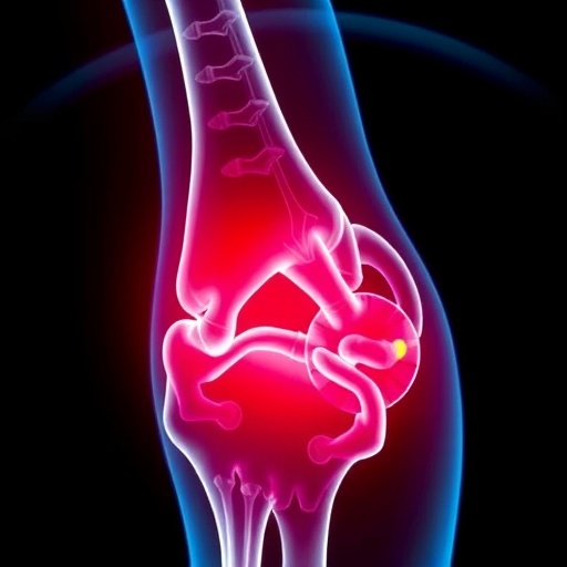
The determination of fracture age is a nuanced and complex endeavor. Traditionally reliant on clinical assessment and radiographic imaging, the field has witnessed a proliferation of advanced imaging technologies over recent decades. Modalities such as conventional radiographs, computed tomography (CT), and magnetic resonance imaging (MRI) present distinct advantages and limitations in different forensic scenarios. Each modality offers varying degrees of sensitivity and specificity, influencing their utility depending on the fracture’s location, patient’s age, and the temporal proximity of injury.
In their comprehensive review, the authors meticulously evaluate how these imaging techniques intersect with specific healing criteria—biological markers observable through imaging that signify the fracture’s progression through stages such as inflammation, soft callus formation, hard callus development, and remodeling. These healing phases carry distinctive radiological signatures that evolve predictably over time but can also be modulated by individual physiological factors and external influences, adding layers of complexity to accurate age estimation.
Radiographic imaging, often the frontline tool in clinical and forensic settings, is dissected with close scrutiny. Plain radiographs provide accessibility and cost-efficiency but are limited by their two-dimensional nature and relatively lower resolution. Findings indicate that while radiographs can effectively identify clear structural changes like callus formation and fracture line visibility, their reliability diminishes when assessing subtle or early healing phases, making them less precise for pinpointing injury timing in complex cases.
Computed tomography (CT) emerges as a superior alternative, offering high-resolution, three-dimensional visualization of bone architecture. The authors highlight CT’s ability to detect minute morphological changes, such as trabecular bone remodeling and cortical bridging, which are critical indicators of healing progression. CT’s enhanced spatial resolution enables forensic experts to discern between fresh and older fractures with greater confidence, although considerations around radiation dose and accessibility remain limiting factors.
Magnetic resonance imaging (MRI), with its exceptional soft tissue contrast, complements CT and radiography by revealing internal biological processes attached to fracture healing, such as marrow edema and vascular changes. This modality provides a unique window into the inflammatory and reparative phases that precede visible ossification. The review underscores MRI’s potential in forensic fracture dating, particularly when the injury is very recent or when accompanying soft tissue damage requires characterization. However, MRI’s utility may be constrained by longer scan times, higher costs, and the requirement for specialized interpretation expertise.
Beyond imaging technologies, the synthesis carefully addresses the healing criteria themselves. Radiographic signs traditionally relied upon include fracture line sharpness, callus opacity, and cortical continuity. The review articulates how temporal patterns of these features, when mapped across multiple studies, can serve as probabilistic markers of fracture age. Notably, the authors stress the influence of confounding factors such as patient age, metabolic conditions, and treatment interventions, which can accelerate or delay healing, thus necessitating a nuanced approach to interpretation.
A critical contribution of this systematic review lies in its comparative evaluation across studies that utilize disparate criteria and imaging modalities. By standardizing terminology and assessment protocols, the authors argue for a harmonization of forensic fracture dating methods. This standardization is vital to improve reproducibility, facilitate meta-analyses, and ultimately guide forensic practitioners toward evidence-based best practices that enhance the objectivity and legal defensibility of their conclusions.
Interdisciplinary collaboration emerges as a key theme in advancing fracture age estimation. The review advocates for integration between radiologists, pathologists, and forensic anthropologists to combine imaging insights with clinical data and histological analyses. Such fusion could yield multi-modal algorithms capable of producing more robust and precise fracture age estimates, delivering impactful advancements in both medico-legal investigations and clinical care.
Underlying this meticulous scientific discourse is the recognition that fracture age estimation is not simply an academic exercise but a tool with profound human and societal implications. Whether resolving disputed timelines in cases of suspected abuse, personal injury, or war crimes, forensic accuracy can alter justice outcomes and provide closure for victims and families. By refining imaging strategies and healing markers, this review propels the forensic community toward greater clarity and reliability.
Looking ahead, the authors encourage future research to leverage emerging technologies such as high-resolution peripheral quantitative CT, advanced MRI sequences, and machine learning approaches. These innovations hold promise for unveiling subtle patterns overlooked by traditional methods. Additionally, the incorporation of longitudinal clinical datasets and experimental models could enhance understanding of variability in healing trajectories, thereby fortifying fracture age assessment frameworks.
This systematic review also serves an educational function, disseminating distilled knowledge to forensic practitioners worldwide. Its detailed analysis empowers experts to critically evaluate the strengths and constraints of their tools, fostering an informed selection of imaging modalities tailored to each investigative context. By elevating methodological rigor, the study contributes to enhanced standards and the overall credibility of forensic science.
In synthesizing decades of heterogeneous research, Lawrence, Williams, and Primeau chart a course toward more precise and nuanced fracture age estimation. Their work exemplifies how systematic analysis can reveal gaps, harmonize criteria, and stimulate innovation — a model for advancing forensic methodologies amid evolving technological landscapes. The forensic community stands on the cusp of significant advancements, fueled by such critical evaluations that merge science, technology, and the pursuit of justice.
As the field progresses, ethical considerations regarding radiation exposure and equitable access to advanced imaging modalities will also demand attention. Balancing scientific rigor with patient and subject welfare underscores the multidisciplinary challenges inherent in forensic fracture dating. These dimensions, woven into the technical discourse, reflect the broader societal responsibilities accompanying scientific innovation.
Ultimately, this systematic review exemplifies the power of comprehensive scholarship combined with technological insight to push the boundaries of forensic science. It invites researchers, clinicians, and legal professionals alike to embrace refined imaging strategies and standardized criteria, fostering a future in which fracture age estimation is marked by precision, reliability, and profound forensic impact.
Subject of Research: Estimation of fracture age through imaging modalities and healing criteria
Article Title: Comparing imaging modalities and healing criteria for the estimation of fracture age: a systematic review
Article References:
Lawrence, C.G., Williams, M.A. & Primeau, C. Comparing imaging modalities and healing criteria for the estimation of fracture age: a systematic review. Int J Legal Med (2025). https://doi.org/10.1007/s00414-025-03589-w
Image Credits: AI Generated
Tags: advanced imaging technologies in medicinebiological markers in fracture healingcomparative efficacy of imaging modalitiesforensic fracture age estimationhealing markers in fracture analysisimaging techniques in forensic sciencejudicial implications of fracture age determinationlegal contexts of fracture analysisrefining forensic methodologies in fracture assessmentrole of CT and MRI in forensicssensitivity and specificity of imaging techniquessystematic review of fracture aging

